

Chapter 10
Resolution in Electron Tomography
Mikhail Kudryashev
Abstract Electron microscopes yield point resolution on the order of one angstrom,
however the density maps from electron tomography typically have resolutions in
the nanometre range. In this chapter I qualitatively discuss the typical limitations that
occur in electron tomography of biological samples depending on the imaging
modalities, with the focus on cryo electron tomography and subtomogram
averaging.
10.1 Introduction
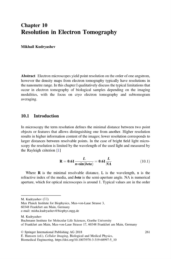
In microscopy the term resolution defines the minimal distance between two point
objects or features that allows distinguishing one from another. Higher resolution
results in higher information content of the images; lower resolution corresponds to
larger distances between resolvable points. In the case of bright field light microscopy the resolution is limited by the wavelength of the used light and measured by
the Rayleigh criterion [1]
R ¼ 0:61
L
nÃsin beta
ð
Þ
¼ 0:61
L
NA
ð10:1Þ
Where R is the minimal resolvable distance, L is the wavelength, n is the
refractive index of the media, and beta is the semi-aperture angle. NA is numerical
aperture, which for optical microscopes is around 1. Typical values are in the order
M. Kudryashev (&)
Max Planck Institute for Biophysics, Max-von-Laue Strasse 3,
60348 Frankfurt am Main, Germany
e-mail: misha.kudryashev@biophys.mpg.de
M. Kudryashev
Buchmann Institute for Molecular Life Sciences, Goethe University
of Frankfurt am Main, Max-von-Laue Strasse 17, 60348 Frankfurt am Main, Germany
© Springer International Publishing AG 2018
E. Hanssen (ed.), Cellular Imaging, Biological and Medical Physics,
Biomedical Engineering, https://doi.org/10.1007/978-3-319-68997-5_10
261
Resolution in Electron Tomography
Mikhail Kudryashev
Abstract Electron microscopes yield point resolution on the order of one angstrom,
however the density maps from electron tomography typically have resolutions in
the nanometre range. In this chapter I qualitatively discuss the typical limitations that
occur in electron tomography of biological samples depending on the imaging
modalities, with the focus on cryo electron tomography and subtomogram
averaging.
10.1 Introduction
In microscopy the term resolution defines the minimal distance between two point
objects or features that allows distinguishing one from another. Higher resolution
results in higher information content of the images; lower resolution corresponds to
larger distances between resolvable points. In the case of bright field light microscopy the resolution is limited by the wavelength of the used light and measured by
the Rayleigh criterion [1]
R ¼ 0:61
L
nÃsin beta
ð
Þ
¼ 0:61
L
NA
ð10:1Þ
Where R is the minimal resolvable distance, L is the wavelength, n is the
refractive index of the media, and beta is the semi-aperture angle. NA is numerical
aperture, which for optical microscopes is around 1. Typical values are in the order
M. Kudryashev (&)
Max Planck Institute for Biophysics, Max-von-Laue Strasse 3,
60348 Frankfurt am Main, Germany
e-mail: misha.kudryashev@biophys.mpg.de
M. Kudryashev
Buchmann Institute for Molecular Life Sciences, Goethe University
of Frankfurt am Main, Max-von-Laue Strasse 17, 60348 Frankfurt am Main, Germany
© Springer International Publishing AG 2018
E. Hanssen (ed.), Cellular Imaging, Biological and Medical Physics,
Biomedical Engineering, https://doi.org/10.1007/978-3-319-68997-5_10
261