

9.6 Available Software
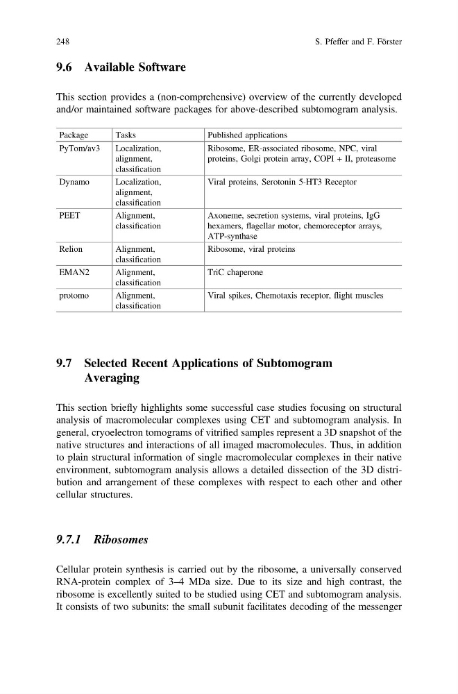
This section provides a (non-comprehensive) overview of the currently developed
and/or maintained software packages for above-described subtomogram analysis.
Package
Tasks
Published applications
PyTom/av3
Localization,
alignment,
classification
Ribosome, ER-associated ribosome, NPC, viral
proteins, Golgi protein array, COPI + II, proteasome
Dynamo
Localization,
alignment,
classification
Viral proteins, Serotonin 5-HT3 Receptor
PEET
Alignment,
classification
Axoneme, secretion systems, viral proteins, IgG
hexamers, flagellar motor, chemoreceptor arrays,
ATP-synthase
Relion
Alignment,
classification
Ribosome, viral proteins
EMAN2
Alignment,
classification
TriC chaperone
protomo
Alignment,
classification
Viral spikes, Chemotaxis receptor, flight muscles
9.7 Selected Recent Applications of Subtomogram
Averaging
This section briefly highlights some successful case studies focusing on structural
analysis of macromolecular complexes using CET and subtomogram analysis. In
general, cryoelectron tomograms of vitrified samples represent a 3D snapshot of the
native structures and interactions of all imaged macromolecules. Thus, in addition
to plain structural information of single macromolecular complexes in their native
environment, subtomogram analysis allows a detailed dissection of the 3D distribution and arrangement of these complexes with respect to each other and other
cellular structures.
9.7.1 Ribosomes
Cellular protein synthesis is carried out by the ribosome, a universally conserved
RNA-protein complex of 3–4 MDa size. Due to its size and high contrast, the
ribosome is excellently suited to be studied using CET and subtomogram analysis.
It consists of two subunits: the small subunit facilitates decoding of the messenger
248
S. Pfeffer and F. Förster
This section provides a (non-comprehensive) overview of the currently developed
and/or maintained software packages for above-described subtomogram analysis.
Package
Tasks
Published applications
PyTom/av3
Localization,
alignment,
classification
Ribosome, ER-associated ribosome, NPC, viral
proteins, Golgi protein array, COPI + II, proteasome
Dynamo
Localization,
alignment,
classification
Viral proteins, Serotonin 5-HT3 Receptor
PEET
Alignment,
classification
Axoneme, secretion systems, viral proteins, IgG
hexamers, flagellar motor, chemoreceptor arrays,
ATP-synthase
Relion
Alignment,
classification
Ribosome, viral proteins
EMAN2
Alignment,
classification
TriC chaperone
protomo
Alignment,
classification
Viral spikes, Chemotaxis receptor, flight muscles
9.7 Selected Recent Applications of Subtomogram
Averaging
This section briefly highlights some successful case studies focusing on structural
analysis of macromolecular complexes using CET and subtomogram analysis. In
general, cryoelectron tomograms of vitrified samples represent a 3D snapshot of the
native structures and interactions of all imaged macromolecules. Thus, in addition
to plain structural information of single macromolecular complexes in their native
environment, subtomogram analysis allows a detailed dissection of the 3D distribution and arrangement of these complexes with respect to each other and other
cellular structures.
9.7.1 Ribosomes
Cellular protein synthesis is carried out by the ribosome, a universally conserved
RNA-protein complex of 3–4 MDa size. Due to its size and high contrast, the
ribosome is excellently suited to be studied using CET and subtomogram analysis.
It consists of two subunits: the small subunit facilitates decoding of the messenger
248
S. Pfeffer and F. Förster