

to generate structures for pleomorphic specimens at a resolution of better than
10 nm. Furthermore, tomograms of cells can be archived and refined locally when
improved structures become available. In the context of cell biology and virology,
this is extremely important: all cells are pleomorphic, and they are subject to
stochastic variability. They may contain symmetrical or repetitive structures, e.g.
icosahedral viruses but tomography is needed to visualise the cellular landscape. On
the other hand, the use of single-particle averaging for determining the structure of
wholly symmetrical viruses is well established and represents a mature technology
[57].
1.3 Sample Preparation for Electron Tomography
The ability of electron tomography to generate authentic structures at a resolution of
better than 10 nm emphasises the requirement for suitable, resolution-preserving
sample preparation procedures. All forms of sample preparation require a fixation
(immobilisation) step to stabilise the structures and maintain their spatial relationships when introduced into the vacuum environment of the electron microscope.
The classical approach is chemical fixation using aldehyde-based cross-linkers,
which are easily criticised as artefacts. Physical immobilisation by rapid freezing
can be used instead, and it is used exclusively for cryo- electron tomography, as
well as for single-particle analysis. In brief, vitrification means that with sufficiently
fast cooling rates, water is transformed into an amorphous solid that is thought to
resemble liquid water, and which is (meta) stable during controlled image acquisition. This is referred to as the ‘frozen-hydrated’ state.
Before discussing the different methods for acquiring, aligning, reconstructing
and segmenting tomograms (see following sections), it is first necessary to mention
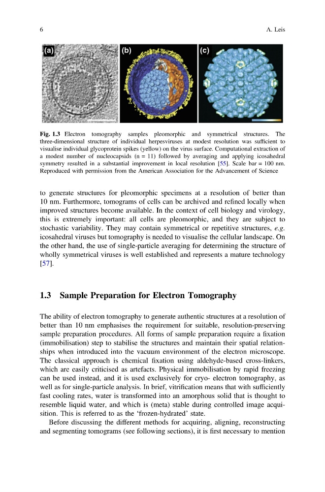
Fig. 1.3 Electron tomography samples pleomorphic and symmetrical structures. The
three-dimensional structure of individual herpesviruses at modest resolution was sufficient to
visualise individual glycoprotein spikes (yellow) on the virus surface. Computational extraction of
a modest number of nucleocapsids (n = 11) followed by averaging and applying icosahedral
symmetry resulted in a substantial improvement in local resolution [55]. Scale bar = 100 nm.
Reproduced with permission from the American Association for the Advancement of Science
6
A. Leis
10 nm. Furthermore, tomograms of cells can be archived and refined locally when
improved structures become available. In the context of cell biology and virology,
this is extremely important: all cells are pleomorphic, and they are subject to
stochastic variability. They may contain symmetrical or repetitive structures, e.g.
icosahedral viruses but tomography is needed to visualise the cellular landscape. On
the other hand, the use of single-particle averaging for determining the structure of
wholly symmetrical viruses is well established and represents a mature technology
[57].
1.3 Sample Preparation for Electron Tomography
The ability of electron tomography to generate authentic structures at a resolution of
better than 10 nm emphasises the requirement for suitable, resolution-preserving
sample preparation procedures. All forms of sample preparation require a fixation
(immobilisation) step to stabilise the structures and maintain their spatial relationships when introduced into the vacuum environment of the electron microscope.
The classical approach is chemical fixation using aldehyde-based cross-linkers,
which are easily criticised as artefacts. Physical immobilisation by rapid freezing
can be used instead, and it is used exclusively for cryo- electron tomography, as
well as for single-particle analysis. In brief, vitrification means that with sufficiently
fast cooling rates, water is transformed into an amorphous solid that is thought to
resemble liquid water, and which is (meta) stable during controlled image acquisition. This is referred to as the ‘frozen-hydrated’ state.
Before discussing the different methods for acquiring, aligning, reconstructing
and segmenting tomograms (see following sections), it is first necessary to mention
Fig. 1.3 Electron tomography samples pleomorphic and symmetrical structures. The
three-dimensional structure of individual herpesviruses at modest resolution was sufficient to
visualise individual glycoprotein spikes (yellow) on the virus surface. Computational extraction of
a modest number of nucleocapsids (n = 11) followed by averaging and applying icosahedral
symmetry resulted in a substantial improvement in local resolution [55]. Scale bar = 100 nm.
Reproduced with permission from the American Association for the Advancement of Science
6
A. Leis