

10 Å (reviewed in [31, 33, 48]). The sample is usually not tilted; exceptions are the
random conical tilt and related orthogonal tilt methods [49]. Instead, the typically
reasonable assumption is made that the macromolecules or viruses lie in random
orientations with respect to the beam. These different views of the ‘same’ structure
contribute more or less equally to the structure solution, and deviations from
similarity become apparent in the ‘class averages’ representing the different orientations. Apart from its similar requirements for a TEM and image processing,
single-particle analysis bears little relationship to the subject of this book, electron
tomography, except that the structures it generates can be used to populate tomograms of cells in the strategy known as ‘visual proteomics’ [1, 38, 50–53]. As such,
it belongs to a separate category of nominally ‘high-resolution’ techniques that
includes X-ray crystallography but also NMR spectroscopy. Another reason for
mentioning it here is that similar computational approaches can be applied to
symmetrical and/or repetitive structures in tomograms to enhance local resolution.
This is known as subtomogram averaging [54], and forms the subject of Chap. 9.
The structure of the Herpes simplex virus (Fig. 1.3) provides an elegant
demonstration of this approach because each mature virus contains both pleomorphic and symmetrical components [55]. The resolution for the symmetrical
components can be enhanced post-acquisition. Thus, even though electron
tomography produces a comparatively modest resolution compared to
single-particle analysis of wholly symmetrical viruses [56], it is unique in its ability
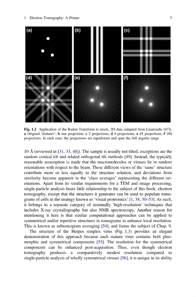
Fig. 1.2 Application of the Radon Transform to mock, 2D data (adapted from Linaroudis [47]).
a Original ‘features’; b one projection; c 2 projections; d 4 projections; e 45 projections; f 180
projections. In each case, the projections are equidistant and span the full angular range
1 Electron Tomography: A Primer
5
random conical tilt and related orthogonal tilt methods [49]. Instead, the typically
reasonable assumption is made that the macromolecules or viruses lie in random
orientations with respect to the beam. These different views of the ‘same’ structure
contribute more or less equally to the structure solution, and deviations from
similarity become apparent in the ‘class averages’ representing the different orientations. Apart from its similar requirements for a TEM and image processing,
single-particle analysis bears little relationship to the subject of this book, electron
tomography, except that the structures it generates can be used to populate tomograms of cells in the strategy known as ‘visual proteomics’ [1, 38, 50–53]. As such,
it belongs to a separate category of nominally ‘high-resolution’ techniques that
includes X-ray crystallography but also NMR spectroscopy. Another reason for
mentioning it here is that similar computational approaches can be applied to
symmetrical and/or repetitive structures in tomograms to enhance local resolution.
This is known as subtomogram averaging [54], and forms the subject of Chap. 9.
The structure of the Herpes simplex virus (Fig. 1.3) provides an elegant
demonstration of this approach because each mature virus contains both pleomorphic and symmetrical components [55]. The resolution for the symmetrical
components can be enhanced post-acquisition. Thus, even though electron
tomography produces a comparatively modest resolution compared to
single-particle analysis of wholly symmetrical viruses [56], it is unique in its ability
Fig. 1.2 Application of the Radon Transform to mock, 2D data (adapted from Linaroudis [47]).
a Original ‘features’; b one projection; c 2 projections; d 4 projections; e 45 projections; f 180
projections. In each case, the projections are equidistant and span the full angular range
1 Electron Tomography: A Primer
5