

pre-processing is not done. Most of this is done in real space, while corrections for
the contrast transfer function is done in frequency space (see next section).
8.4.1 Normalizing Images
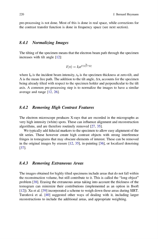
The tilting of the specimen means that the electron beam path through the specimen
increases with tilt angle [12]:
IðaÞ ¼ I 0 e
t 0
K cosða þ DaÞ
where I 0 is the incident beam intensity, t 0 is the specimen thickness at zero-tilt, and
K is the mean free path. The addition to the tilt angle, Δa, accounts for the specimen
being already tilted with respect to the specimen holder and perpendicular to the tilt
axis. A common pre-processing step is to normalize the images to have a similar
average and range [12, 26].
8.4.2 Removing High Contrast Features
The electron microscope produces X-rays that are recorded in the micrographs as
very high intensity (white) spots. These can influence alignment and reconstruction
algorithms, and are therefore routinely removed [27, 35].
We typically add fiducial markers to the specimen to allow easy alignment of the
tilt series. These however create high contrast objects with strong interference
fringes in tomograms that may obscure elements of interest. These can be removed
in the original images by erasure [12, 35], in-painting [36], or localized denoising
[37].
8.4.3 Removing Extraneous Areas
The images obtained for highly tilted specimens include areas that do not fall within
the reconstruction volume, but still contribute to it. This is called the “long object”
problem [38]. Erasing the extraneous areas taking into account the thickness of the
tomogram can minimize their contributions (implemented as an option in Bsoft
[12]). Xu et al. [39] incorporated a scheme to weigh down these areas during SIRT.
Turoň ová et al. [40] suggested other ways of dealing with it, including larger
reconstructions to include the additional areas, and appropriate weighting.
220
J. Bernard Heymann
the contrast transfer function is done in frequency space (see next section).
8.4.1 Normalizing Images
The tilting of the specimen means that the electron beam path through the specimen
increases with tilt angle [12]:
IðaÞ ¼ I 0 e
t 0
K cosða þ DaÞ
where I 0 is the incident beam intensity, t 0 is the specimen thickness at zero-tilt, and
K is the mean free path. The addition to the tilt angle, Δa, accounts for the specimen
being already tilted with respect to the specimen holder and perpendicular to the tilt
axis. A common pre-processing step is to normalize the images to have a similar
average and range [12, 26].
8.4.2 Removing High Contrast Features
The electron microscope produces X-rays that are recorded in the micrographs as
very high intensity (white) spots. These can influence alignment and reconstruction
algorithms, and are therefore routinely removed [27, 35].
We typically add fiducial markers to the specimen to allow easy alignment of the
tilt series. These however create high contrast objects with strong interference
fringes in tomograms that may obscure elements of interest. These can be removed
in the original images by erasure [12, 35], in-painting [36], or localized denoising
[37].
8.4.3 Removing Extraneous Areas
The images obtained for highly tilted specimens include areas that do not fall within
the reconstruction volume, but still contribute to it. This is called the “long object”
problem [38]. Erasing the extraneous areas taking into account the thickness of the
tomogram can minimize their contributions (implemented as an option in Bsoft
[12]). Xu et al. [39] incorporated a scheme to weigh down these areas during SIRT.
Turoň ová et al. [40] suggested other ways of dealing with it, including larger
reconstructions to include the additional areas, and appropriate weighting.
220
J. Bernard Heymann