

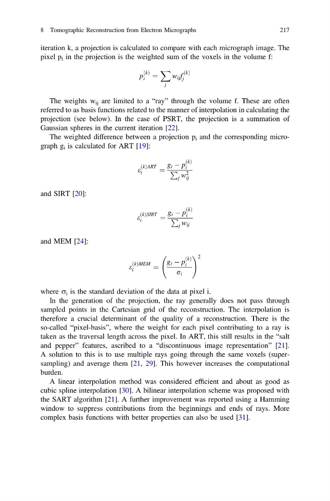
iteration k, a projection is calculated to compare with each micrograph image. The
pixel p i in the projection is the weighted sum of the voxels in the volume f:
p
ðkÞ
i ¼
X
j
w ij f
ðkÞ
j
The weights w ij are limited to a “ray” through the volume f. These are often
referred to as basis functions related to the manner of interpolation in calculating the
projection (see below). In the case of PSRT, the projection is a summation of
Gaussian spheres in the current iteration [22].
The weighted difference between a projection p i and the corresponding micrograph g i is calculated for ART [19]:
e
ðkÞART
i
¼
g i À p
ðkÞ
i
P
j w 2
ij
and SIRT [20]:
e
ðkÞSIRT
i
¼
g i À p
ðkÞ
i
P
j w ij
and MEM [24]:
e
ðkÞMEM
i
¼
g i À p
ðkÞ
i
r i
! 2
where r i is the standard deviation of the data at pixel i.
In the generation of the projection, the ray generally does not pass through
sampled points in the Cartesian grid of the reconstruction. The interpolation is
therefore a crucial determinant of the quality of a reconstruction. There is the
so-called “pixel-basis”, where the weight for each pixel contributing to a ray is
taken as the traversal length across the pixel. In ART, this still results in the “salt
and pepper” features, ascribed to a “discontinuous image representation” [21].
A solution to this is to use multiple rays going through the same voxels (supersampling) and average them [21, 29]. This however increases the computational
burden.
A linear interpolation method was considered efficient and about as good as
cubic spline interpolation [30]. A bilinear interpolation scheme was proposed with
the SART algorithm [21]. A further improvement was reported using a Hamming
window to suppress contributions from the beginnings and ends of rays. More
complex basis functions with better properties can also be used [31].
8 Tomographic Reconstruction from Electron Micrographs
217
pixel p i in the projection is the weighted sum of the voxels in the volume f:
p
ðkÞ
i ¼
X
j
w ij f
ðkÞ
j
The weights w ij are limited to a “ray” through the volume f. These are often
referred to as basis functions related to the manner of interpolation in calculating the
projection (see below). In the case of PSRT, the projection is a summation of
Gaussian spheres in the current iteration [22].
The weighted difference between a projection p i and the corresponding micrograph g i is calculated for ART [19]:
e
ðkÞART
i
¼
g i À p
ðkÞ
i
P
j w 2
ij
and SIRT [20]:
e
ðkÞSIRT
i
¼
g i À p
ðkÞ
i
P
j w ij
and MEM [24]:
e
ðkÞMEM
i
¼
g i À p
ðkÞ
i
r i
! 2
where r i is the standard deviation of the data at pixel i.
In the generation of the projection, the ray generally does not pass through
sampled points in the Cartesian grid of the reconstruction. The interpolation is
therefore a crucial determinant of the quality of a reconstruction. There is the
so-called “pixel-basis”, where the weight for each pixel contributing to a ray is
taken as the traversal length across the pixel. In ART, this still results in the “salt
and pepper” features, ascribed to a “discontinuous image representation” [21].
A solution to this is to use multiple rays going through the same voxels (supersampling) and average them [21, 29]. This however increases the computational
burden.
A linear interpolation method was considered efficient and about as good as
cubic spline interpolation [30]. A bilinear interpolation scheme was proposed with
the SART algorithm [21]. A further improvement was reported using a Hamming
window to suppress contributions from the beginnings and ends of rays. More
complex basis functions with better properties can also be used [31].
8 Tomographic Reconstruction from Electron Micrographs
217