

together the connectome, but has since moved to working on an extensive range of
biological samples (see [16–18]). The technique has also been used in material
science research [19, 20] though its uptake there has been slower.
In contrast to SBEM, the FIB-SEM was developed for use in materials science
and nano-technology applications [21]. The first use of a liquid metal ion source
(LMIS) was documented by Krohn [22], who was trying to develop thrusters for
use in space. In the 1980’s, FIB systems based on high-brightness gallium LMISs
became commercially available [23]. One of the first FIB applications in life sciences was Yonehara et al. [24], where they used an argon ion beam to mill biological specimens then observed with a low accelerating voltage SEM in the same
(a)
(d)
(e)
(b)
(c)
(g)
(h)
(f)
T
S
K
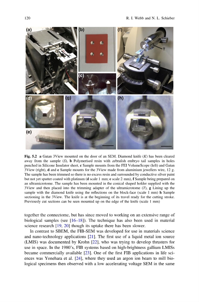
Fig. 5.2 a Gatan 3View mounted on the door of an SEM. Diamond knife (K) has been cleared
away from the sample (S), b Polymerised resin with zebrafish embryo tail samples in holes
punched in Silicone Insulator sheet, c Sample mounts from the FEI VolumeScope (left) and Gatan
3View (right), d and e Sample mounts for the 3View made from aluminium jewellers wire, 12 g.
The sample has been trimmed so there is no excess resin and surrounded by conductive silver paint
but not yet sputter coated with platinum (d scale 1 mm; e scale 2 mm), f Sample being prepared on
an ultramicrotome. The sample has been mounted in the conical shaped holder supplied with the
3View and then placed into the trimming adapter of the ultramicrotome (T), g Lining up the
sample with the diamond knife using the reflections on the block-face (scale 1 mm) h Sample
sectioning in the 3View. The knife is at the beginning of its travel ready for the cutting stroke.
Previously cut sections can be seen mounted up on the edge of the knife (scale 1 mm)
120
R. I. Webb and N. L. Schieber
biological samples (see [16–18]). The technique has also been used in material
science research [19, 20] though its uptake there has been slower.
In contrast to SBEM, the FIB-SEM was developed for use in materials science
and nano-technology applications [21]. The first use of a liquid metal ion source
(LMIS) was documented by Krohn [22], who was trying to develop thrusters for
use in space. In the 1980’s, FIB systems based on high-brightness gallium LMISs
became commercially available [23]. One of the first FIB applications in life sciences was Yonehara et al. [24], where they used an argon ion beam to mill biological specimens then observed with a low accelerating voltage SEM in the same
(a)
(d)
(e)
(b)
(c)
(g)
(h)
(f)
T
S
K
Fig. 5.2 a Gatan 3View mounted on the door of an SEM. Diamond knife (K) has been cleared
away from the sample (S), b Polymerised resin with zebrafish embryo tail samples in holes
punched in Silicone Insulator sheet, c Sample mounts from the FEI VolumeScope (left) and Gatan
3View (right), d and e Sample mounts for the 3View made from aluminium jewellers wire, 12 g.
The sample has been trimmed so there is no excess resin and surrounded by conductive silver paint
but not yet sputter coated with platinum (d scale 1 mm; e scale 2 mm), f Sample being prepared on
an ultramicrotome. The sample has been mounted in the conical shaped holder supplied with the
3View and then placed into the trimming adapter of the ultramicrotome (T), g Lining up the
sample with the diamond knife using the reflections on the block-face (scale 1 mm) h Sample
sectioning in the 3View. The knife is at the beginning of its travel ready for the cutting stroke.
Previously cut sections can be seen mounted up on the edge of the knife (scale 1 mm)
120
R. I. Webb and N. L. Schieber