

while SEM relies on a raster scan of the sample, which can also limit the resolution.
In the TEM, images are formed from 2D projected information coming from the full
depth of the section (50 nm) resulting in the overlaping of structures. Volume
SEMs collect backscattered electrons coming from a limited depth within the
sample (dependent on the voltage but usually only a few nanometres), resulting in a
better Z resolved image.
The concept of placing a microtome into an SEM so that the sample could be
imaged sequentially as pieces of it were removed was first conceived by Leighton in
1981 [12]. It was then fully developed by Denk and Horstmann [5] and Gatan
released a commercialised version (3View) years later [5]. The 3View (Fig. 5.2a),
can be installed onto an SEM from several manufacturers: FEI, Zeiss, JEOL,
Hitachi and Tescan. In 2015, FEI released their own version, the VolumeScope, on
their Teneo SEM as a fully integrated package. Along with this they released a
deconvolution system [13–15], which captures images using different landing
energies after each section. By deconvolving the information from multiple images
it is possible to obtain 10 nm isotropic resolution using this SBEM. Initially the
SBEM technology was widely used in neurobiological studies in order to piece
Serial Block-Face Scanning Electron Microscopy (SBEM)
Milling
Sectioning
Sample
Sample
Electron
Beam
Electron
Beam
SEM
SEM
Focused
Ion Beam
SEM
SEM
Diamond
Knife
Imaging
Focused Ion Beam Scanning Electron Microscopy (FIB-SEM)
Imaging
Sample
Sample
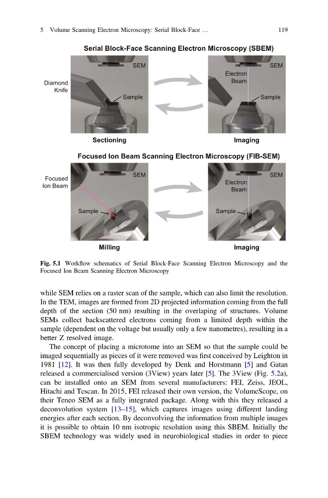
Fig. 5.1 Workflow schematics of Serial Block-Face Scanning Electron Microscopy and the
Focused Ion Beam Scanning Electron Microscopy
5 Volume Scanning Electron Microscopy: Serial Block-Face …
119
In the TEM, images are formed from 2D projected information coming from the full
depth of the section (50 nm) resulting in the overlaping of structures. Volume
SEMs collect backscattered electrons coming from a limited depth within the
sample (dependent on the voltage but usually only a few nanometres), resulting in a
better Z resolved image.
The concept of placing a microtome into an SEM so that the sample could be
imaged sequentially as pieces of it were removed was first conceived by Leighton in
1981 [12]. It was then fully developed by Denk and Horstmann [5] and Gatan
released a commercialised version (3View) years later [5]. The 3View (Fig. 5.2a),
can be installed onto an SEM from several manufacturers: FEI, Zeiss, JEOL,
Hitachi and Tescan. In 2015, FEI released their own version, the VolumeScope, on
their Teneo SEM as a fully integrated package. Along with this they released a
deconvolution system [13–15], which captures images using different landing
energies after each section. By deconvolving the information from multiple images
it is possible to obtain 10 nm isotropic resolution using this SBEM. Initially the
SBEM technology was widely used in neurobiological studies in order to piece
Serial Block-Face Scanning Electron Microscopy (SBEM)
Milling
Sectioning
Sample
Sample
Electron
Beam
Electron
Beam
SEM
SEM
Focused
Ion Beam
SEM
SEM
Diamond
Knife
Imaging
Focused Ion Beam Scanning Electron Microscopy (FIB-SEM)
Imaging
Sample
Sample
Fig. 5.1 Workflow schematics of Serial Block-Face Scanning Electron Microscopy and the
Focused Ion Beam Scanning Electron Microscopy
5 Volume Scanning Electron Microscopy: Serial Block-Face …
119