

6. Make that image a new map by pressing the New Map button in the Navigator
window dialogue.
7. Go to a lower magnification where the field of view is at least 10 µm (typically *4000Â) and take a Record image without moving the stage.
8. Save the image in the same file.
9. Make that image a New Map.
10. Check “For anchor state” on the lower magnification image in the Navigator
window.
11. Go to a new area of interest or find the area/cell on an adjacent section.
12. Take a Record image at the tilt series magnification.
13. Save the image.
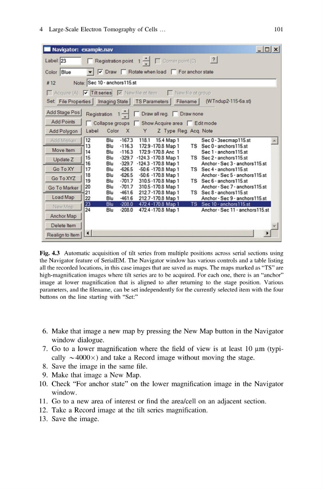
Fig. 4.3 Automatic acquisition of tilt series from multiple positions across serial sections using
the Navigator feature of SerialEM. The Navigator window has various controls and a table listing
all the recorded locations, in this case images that are saved as maps. The maps marked as “TS” are
high-magnification images where tilt series are to be acquired. For each one, there is an “anchor”
image at lower magnification that is aligned to after returning to the stage position. Various
parameters, and the filename, can be set independently for the currently selected item with the four
buttons on the line starting with “Set:”
4 Large-Scale Electron Tomography of Cells …
101
window dialogue.
7. Go to a lower magnification where the field of view is at least 10 µm (typically *4000Â) and take a Record image without moving the stage.
8. Save the image in the same file.
9. Make that image a New Map.
10. Check “For anchor state” on the lower magnification image in the Navigator
window.
11. Go to a new area of interest or find the area/cell on an adjacent section.
12. Take a Record image at the tilt series magnification.
13. Save the image.
Fig. 4.3 Automatic acquisition of tilt series from multiple positions across serial sections using
the Navigator feature of SerialEM. The Navigator window has various controls and a table listing
all the recorded locations, in this case images that are saved as maps. The maps marked as “TS” are
high-magnification images where tilt series are to be acquired. For each one, there is an “anchor”
image at lower magnification that is aligned to after returning to the stage position. Various
parameters, and the filename, can be set independently for the currently selected item with the four
buttons on the line starting with “Set:”
4 Large-Scale Electron Tomography of Cells …
101