

channel ends [117]. Selective amino functionalization of the channel entrances of ZL
crystals, which were loaded with Py
+ as luminescent label, was realized using the
reaction procedure explained in Fig. 7b [87]. The bacteria-{Py
+
-ZL} assembly was
observed to be stable, presumably owing to electrostatic interactions. Fluorescence
microscopy was employed in solution to characterize the assembly. The Py
+
-ZL can
be easily detected in the aggregate, because of its characteristic green luminescence.
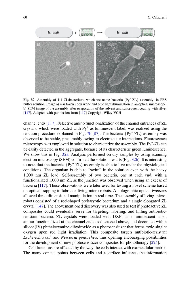
We show this in Fig. 32a. Analysis performed on dry samples by using scanning
electron microscopy (SEM) confirmed the solution results (Fig. 32b). It is interesting
to note that the bacteria-{Py
+
-ZL} assembly is able to live under the physiological
conditions. The organism is able to “swim” in the solution even with the heavy
1,000 nm ZL load. Self-assembly of two bacteria, one at each end, with a
functionalized 1,000 nm ZL as the junction was observed when using an excess of
bacteria [117]. These observations were later used for testing a novel scheme based
on optical trapping to fabricate living micro-robots. A holographic optical tweezers
allowed three-dimensional manipulation in real time. The assembly of living microrobots consisted of a rod-shaped prokaryotic bacterium and a single elongated ZL
crystal [147]. The abovementioned discovery was also used to test if photoactive ZL
composites could eventually serve for targeting, labeling, and killing antibioticresistant bacteria. ZL crystals were loaded with DXP, as a luminescent label,
amino functionalized at the channel ends as discussed above, and decorated with a
silicon(IV) phthalocyanine dihydroxide as a photosensitizer that forms toxic singlet
oxygen upon red light irradiation. This composite targets antibiotic-resistant
Escherichia coli and Neisseria gonorrhea, thus opening encouraging possibilities
for the development of new photosensitizer composites for phototherapy [224].
Cell functions are affected by the way the cells interact with extracellular matrix.
The many contact points between cells and a surface influence the information
Fig. 32 Assembly of 1:1 ZL/bacterium, which we name bacteria-{Py
+ -ZL} assembly, in PBS
buffer solution. Image a) was taken upon white and blue light illumination in an optical microscope.
b) SEM image of the assembly after evaporation of the solvent and subsequent coating with silver
[117]. Adapted with permission from [117] Copyright Wiley VCH
60
G. Calzaferri
crystals, which were loaded with Py
+ as luminescent label, was realized using the
reaction procedure explained in Fig. 7b [87]. The bacteria-{Py
+
-ZL} assembly was
observed to be stable, presumably owing to electrostatic interactions. Fluorescence
microscopy was employed in solution to characterize the assembly. The Py
+
-ZL can
be easily detected in the aggregate, because of its characteristic green luminescence.
We show this in Fig. 32a. Analysis performed on dry samples by using scanning
electron microscopy (SEM) confirmed the solution results (Fig. 32b). It is interesting
to note that the bacteria-{Py
+
-ZL} assembly is able to live under the physiological
conditions. The organism is able to “swim” in the solution even with the heavy
1,000 nm ZL load. Self-assembly of two bacteria, one at each end, with a
functionalized 1,000 nm ZL as the junction was observed when using an excess of
bacteria [117]. These observations were later used for testing a novel scheme based
on optical trapping to fabricate living micro-robots. A holographic optical tweezers
allowed three-dimensional manipulation in real time. The assembly of living microrobots consisted of a rod-shaped prokaryotic bacterium and a single elongated ZL
crystal [147]. The abovementioned discovery was also used to test if photoactive ZL
composites could eventually serve for targeting, labeling, and killing antibioticresistant bacteria. ZL crystals were loaded with DXP, as a luminescent label,
amino functionalized at the channel ends as discussed above, and decorated with a
silicon(IV) phthalocyanine dihydroxide as a photosensitizer that forms toxic singlet
oxygen upon red light irradiation. This composite targets antibiotic-resistant
Escherichia coli and Neisseria gonorrhea, thus opening encouraging possibilities
for the development of new photosensitizer composites for phototherapy [224].
Cell functions are affected by the way the cells interact with extracellular matrix.
The many contact points between cells and a surface influence the information
Fig. 32 Assembly of 1:1 ZL/bacterium, which we name bacteria-{Py
+ -ZL} assembly, in PBS
buffer solution. Image a) was taken upon white and blue light illumination in an optical microscope.
b) SEM image of the assembly after evaporation of the solvent and subsequent coating with silver
[117]. Adapted with permission from [117] Copyright Wiley VCH
60
G. Calzaferri