

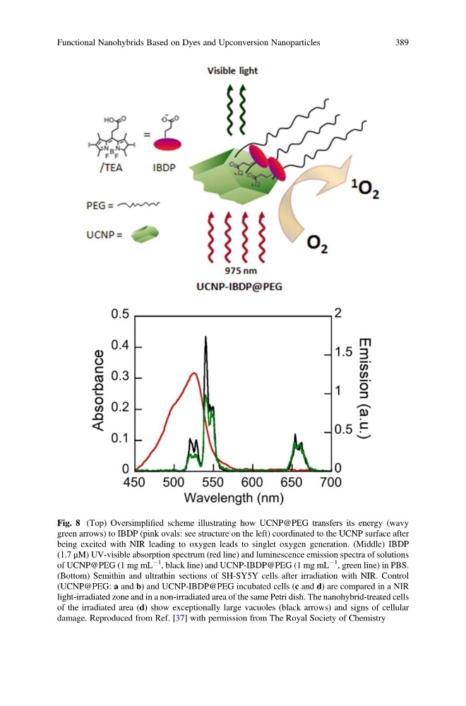
Fig. 8 (Top) Oversimplified scheme illustrating how UCNP@PEG transfers its energy (wavy
green arrows) to IBDP (pink ovals: see structure on the left) coordinated to the UCNP surface after
being excited with NIR leading to oxygen leads to singlet oxygen generation. (Middle) IBDP
(1.7 μM) UV-visible absorption spectrum (red line) and luminescence emission spectra of solutions
of UCNP@PEG (1 mg mL
À1
, black line) and UCNP-IBDP@PEG (1 mg mL
À1
, green line) in PBS.
(Bottom) Semithin and ultrathin sections of SH-SY5Y cells after irradiation with NIR. Control
(UCNP@PEG: a and b) and UCNP-IBDP@PEG incubated cells (c and d) are compared in a NIR
light-irradiated zone and in a non-irradiated area of the same Petri dish. The nanohybrid-treated cells
of the irradiated area (d) show exceptionally large vacuoles (black arrows) and signs of cellular
damage. Reproduced from Ref. [37] with permission from The Royal Society of Chemistry
Functional Nanohybrids Based on Dyes and Upconversion Nanoparticles
389
green arrows) to IBDP (pink ovals: see structure on the left) coordinated to the UCNP surface after
being excited with NIR leading to oxygen leads to singlet oxygen generation. (Middle) IBDP
(1.7 μM) UV-visible absorption spectrum (red line) and luminescence emission spectra of solutions
of UCNP@PEG (1 mg mL
À1
, black line) and UCNP-IBDP@PEG (1 mg mL
À1
, green line) in PBS.
(Bottom) Semithin and ultrathin sections of SH-SY5Y cells after irradiation with NIR. Control
(UCNP@PEG: a and b) and UCNP-IBDP@PEG incubated cells (c and d) are compared in a NIR
light-irradiated zone and in a non-irradiated area of the same Petri dish. The nanohybrid-treated cells
of the irradiated area (d) show exceptionally large vacuoles (black arrows) and signs of cellular
damage. Reproduced from Ref. [37] with permission from The Royal Society of Chemistry
Functional Nanohybrids Based on Dyes and Upconversion Nanoparticles
389