

The vascular endothelial growth factor (VEGF) is an important biomarker for
cancer diagnostics as well as for other diseases [48, 49]. Secreted into the blood for
the purpose of angiogenesis, healthy individuals have serum concentrations of
1–177 pg/ml VEGF while individuals with cancer show a serum level of
18–328 pg/ml VEGF due to increased tissue formation [50]. Therefore, highly
sensitive detection methods are required in order to detect the fine differences
between healthy and cancerous VEGF blood concentrations [51].
In 2015, two studies were published presenting impedimetric aptasensors for
VEGF detection in human serum. Tabrizi et al. reached a linear range of 10–300 pg/
ml and a detection limit of 1 pg/ml by using mesoporous gold nanocomposites on a
screen-printed electrode [49]. For the immobilization, the affinity of gold toward
thiolated aptamer molecules was used. As proof for a potential application, diluted
serum of a patient with lung cancer was tested. The value measured with a standard
ELISA at the local hospital could be recovered to 97% by the developed biosensor.
A more exotic approach was proposed by Qureshi et al. who designed a
non-faradaic aptasensor. Instead of impedance change, this sensor used changes in
capacitance [48]. A linear detection range of 400 pg/ml up to 1 ng/ml VEGF in
spiked serum was obtained for a low volume sample of 5 μl (LoD: 5 pg/ml). The
authors showed the increased capacitance change by a sandwich assay design
compared to a simple aptamer immobilized on a gold surface. The sandwich assay
additionally included an antibody linked to a magnetic bead (see Fig. 5). The
approach is promising with regard to potential parallelization and the need of low
volumes. However, the complexity of the approach might be limiting due to the need
of an additional element such as an eventually expensive antibody. Moreover, the
antibody must not bind the same epitope as the aptamer. The approaches presented
by Tabrizi et al. and Qureshi et al. are close to the medically relevant range.
Compared to other VEGF-biosensors, the presented impedimetric biosensors
stand out due to its high sensitivity even in a complex biological matrix (see
Table 4). Da et al. presented an aptasensor based on photoelectrochemical detection
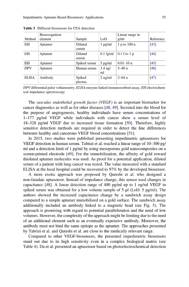
Table 3 Different biosensors for CEA detection
Method
Biorecognition
element
Sample
LoD
Linear range in
g/ml
Reference
EIS
Aptamer
Diluted
serum
1 pg/ml
1 p to 100 n
[43]
EIS
Aptamer
Diluted
serum
0.1 fg/ml 0.1 f to 1 p
[44]
EIS
Aptamer
Spiked serum 5 pg/ml
0.01–10 n
[45]
DPV
Aptamer
Human serum 3.4 ng/
ml
5–40 n
[46]
ELISA Antibody
Spiked
plasma
2 ng/ml
2–64 n
[47]
DPV differential pulse voltammetry, ELISA enzyme-linked immunosorbent assay, EIS electrochemical impedance spectroscopy
Impedimetric Aptamer-Based Biosensors: Applications
55
cancer diagnostics as well as for other diseases [48, 49]. Secreted into the blood for
the purpose of angiogenesis, healthy individuals have serum concentrations of
1–177 pg/ml VEGF while individuals with cancer show a serum level of
18–328 pg/ml VEGF due to increased tissue formation [50]. Therefore, highly
sensitive detection methods are required in order to detect the fine differences
between healthy and cancerous VEGF blood concentrations [51].
In 2015, two studies were published presenting impedimetric aptasensors for
VEGF detection in human serum. Tabrizi et al. reached a linear range of 10–300 pg/
ml and a detection limit of 1 pg/ml by using mesoporous gold nanocomposites on a
screen-printed electrode [49]. For the immobilization, the affinity of gold toward
thiolated aptamer molecules was used. As proof for a potential application, diluted
serum of a patient with lung cancer was tested. The value measured with a standard
ELISA at the local hospital could be recovered to 97% by the developed biosensor.
A more exotic approach was proposed by Qureshi et al. who designed a
non-faradaic aptasensor. Instead of impedance change, this sensor used changes in
capacitance [48]. A linear detection range of 400 pg/ml up to 1 ng/ml VEGF in
spiked serum was obtained for a low volume sample of 5 μl (LoD: 5 pg/ml). The
authors showed the increased capacitance change by a sandwich assay design
compared to a simple aptamer immobilized on a gold surface. The sandwich assay
additionally included an antibody linked to a magnetic bead (see Fig. 5). The
approach is promising with regard to potential parallelization and the need of low
volumes. However, the complexity of the approach might be limiting due to the need
of an additional element such as an eventually expensive antibody. Moreover, the
antibody must not bind the same epitope as the aptamer. The approaches presented
by Tabrizi et al. and Qureshi et al. are close to the medically relevant range.
Compared to other VEGF-biosensors, the presented impedimetric biosensors
stand out due to its high sensitivity even in a complex biological matrix (see
Table 4). Da et al. presented an aptasensor based on photoelectrochemical detection
Table 3 Different biosensors for CEA detection
Method
Biorecognition
element
Sample
LoD
Linear range in
g/ml
Reference
EIS
Aptamer
Diluted
serum
1 pg/ml
1 p to 100 n
[43]
EIS
Aptamer
Diluted
serum
0.1 fg/ml 0.1 f to 1 p
[44]
EIS
Aptamer
Spiked serum 5 pg/ml
0.01–10 n
[45]
DPV
Aptamer
Human serum 3.4 ng/
ml
5–40 n
[46]
ELISA Antibody
Spiked
plasma
2 ng/ml
2–64 n
[47]
DPV differential pulse voltammetry, ELISA enzyme-linked immunosorbent assay, EIS electrochemical impedance spectroscopy
Impedimetric Aptamer-Based Biosensors: Applications
55