

targeted cells could be detached from a cell culture surface by rapid changes of the
direction of the magnetic field. This method might also be useable in the treatment of
solid tumors. Additionally the target cells can be destroyed by the nanosurgeons
presumably caused by mechanical strain-induced apoptosis or necrosis. Therefore
aptamer-modified superparamagnetic nanoparticles are a promising tool for surgical
actions on cellular level [114]. Table 6 shows the compounds of the nanosurgeon
system developed by Nair et al.
Aptamer-Modified Gold Nanoparticles for Reversible Treatment
of Blood-Clotting Disorders
Another extraordinary method that uses an aptamer-modified nanoparticle system
was developed by Huang et al. They used modified aptamers as drugs in combination with gold nanoparticles. These triblock aptamers contained a poly A sequence
for self-assembly on the surface of the gold nanoparticles, a short sequence that can
be used for hybridization and a thrombin binding block. Two different triblock
aptamers were used which contain two thrombin binding aptamers that bind to
different parts of thrombin. For drug preparation both triblock aptamers were
hybridized and then immobilized to gold nanoparticles (they build a self-assembled
monolayer on the nanoparticle surface). Due to the spatial proximity of the two
aptamers, they can bind thrombin effectively and thus prevent the conversion of
fibrin into fibrinogen and further prevent blood clotting. In emergencies, where
blood clotting is required, the aptamers can be detached from the gold nanoparticles
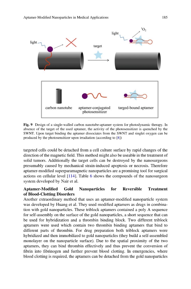
Fig. 9 Design of a single-walled carbon nanotube-aptamer system for photodynamic therapy. In
absence of the target of the used aptamer, the activity of the photosensitizer is quenched by the
SWNT. Upon target binding the aptamer dissociates from the SWNT and singlet oxygen can be
produced by the photosensitizer upon irradiation (according to [8])
Aptamer-Modified Nanoparticles in Medical Applications
185
direction of the magnetic field. This method might also be useable in the treatment of
solid tumors. Additionally the target cells can be destroyed by the nanosurgeons
presumably caused by mechanical strain-induced apoptosis or necrosis. Therefore
aptamer-modified superparamagnetic nanoparticles are a promising tool for surgical
actions on cellular level [114]. Table 6 shows the compounds of the nanosurgeon
system developed by Nair et al.
Aptamer-Modified Gold Nanoparticles for Reversible Treatment
of Blood-Clotting Disorders
Another extraordinary method that uses an aptamer-modified nanoparticle system
was developed by Huang et al. They used modified aptamers as drugs in combination with gold nanoparticles. These triblock aptamers contained a poly A sequence
for self-assembly on the surface of the gold nanoparticles, a short sequence that can
be used for hybridization and a thrombin binding block. Two different triblock
aptamers were used which contain two thrombin binding aptamers that bind to
different parts of thrombin. For drug preparation both triblock aptamers were
hybridized and then immobilized to gold nanoparticles (they build a self-assembled
monolayer on the nanoparticle surface). Due to the spatial proximity of the two
aptamers, they can bind thrombin effectively and thus prevent the conversion of
fibrin into fibrinogen and further prevent blood clotting. In emergencies, where
blood clotting is required, the aptamers can be detached from the gold nanoparticles
Fig. 9 Design of a single-walled carbon nanotube-aptamer system for photodynamic therapy. In
absence of the target of the used aptamer, the activity of the photosensitizer is quenched by the
SWNT. Upon target binding the aptamer dissociates from the SWNT and singlet oxygen can be
produced by the photosensitizer upon irradiation (according to [8])
Aptamer-Modified Nanoparticles in Medical Applications
185