

half-times for dissociation [69], compared to the most stable protein alphahelices [70].
The recognition functionality of an aptamer used as chromatographic ligand is
only marginally dependent on the linear sequence of the nuclear bases, but more
importantly on the organization of double-helical regions, as well as the highly
folded single-stranded stretches. This complex configuration is at the basis of
complementary molecular interactions on given protein aptatopes, a molecular
recognition that is fundamental in affinity chromatography.
While comparing the process allowing the formation of a tertiary structure
between a nucleic acid and a protein, it can be said that the only common driving
force is to minimize the exposure to water. In fact, in the first case, the formation of a
compact structure is determined by the stacking interaction between complementary
bases, while for proteins, the collapse of the structure is mainly due to the formation
of hydrophobic cores. Similarities and differences of secondary and tertiary structure
modeling between proteins and nucleic acids have been recently reviewed [71].
Even in the presence of relatively short oligonucleotide sequences (e.g., tRNA
and also aptamers), it is not an easy task to predict the secondary structure. Most
generally, these structures are reached in order to minimize the level of free energy;
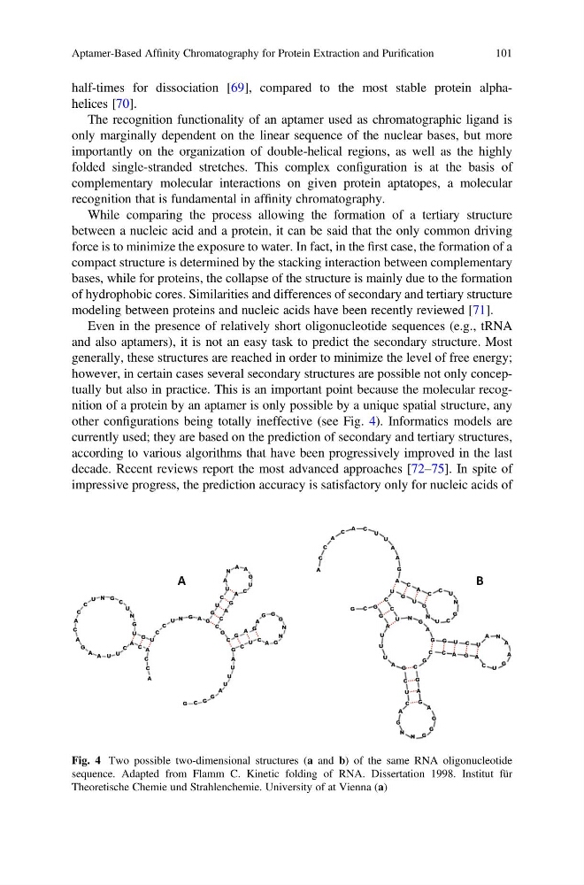
however, in certain cases several secondary structures are possible not only conceptually but also in practice. This is an important point because the molecular recognition of a protein by an aptamer is only possible by a unique spatial structure, any
other configurations being totally ineffective (see Fig. 4). Informatics models are
currently used; they are based on the prediction of secondary and tertiary structures,
according to various algorithms that have been progressively improved in the last
decade. Recent reviews report the most advanced approaches [72–75]. In spite of
impressive progress, the prediction accuracy is satisfactory only for nucleic acids of
A
A
A
A
A
A
A
A
A A
A
A
C
C
C
C
C
C
C
C
C
U
U
U
U
U
U
U
U
U
U
U
U
U
U
G
G
G
G
G
G
G
G
G
A
G
G
G
G
G
G
N
N
N
N
N
N
C
C
C
C
C
C
C
C
C
A
A
A
A
A
G
G
G
G
G
A
A
C
C
C
C
C
C
C
G
G
G
A
U
U
U
U
A
A
A
A
G
G
G
C
C
C
C
C
C
C
C
C
C
U
U
U
U
U
U
U
U
U
U
G
G
G
G
G
G
G
G
G
N N
N
N
N
N
A
A
A
A
A
A
A
A
A
A
A
C
G
G
G
G
G
B
A
Fig. 4 Two possible two-dimensional structures (a and b) of the same RNA oligonucleotide
sequence. Adapted from Flamm C. Kinetic folding of RNA. Dissertation 1998. Institut für
Theoretische Chemie und Strahlenchemie. University of at Vienna (a)
Aptamer-Based Affinity Chromatography for Protein Extraction and Purification
101
The recognition functionality of an aptamer used as chromatographic ligand is
only marginally dependent on the linear sequence of the nuclear bases, but more
importantly on the organization of double-helical regions, as well as the highly
folded single-stranded stretches. This complex configuration is at the basis of
complementary molecular interactions on given protein aptatopes, a molecular
recognition that is fundamental in affinity chromatography.
While comparing the process allowing the formation of a tertiary structure
between a nucleic acid and a protein, it can be said that the only common driving
force is to minimize the exposure to water. In fact, in the first case, the formation of a
compact structure is determined by the stacking interaction between complementary
bases, while for proteins, the collapse of the structure is mainly due to the formation
of hydrophobic cores. Similarities and differences of secondary and tertiary structure
modeling between proteins and nucleic acids have been recently reviewed [71].
Even in the presence of relatively short oligonucleotide sequences (e.g., tRNA
and also aptamers), it is not an easy task to predict the secondary structure. Most
generally, these structures are reached in order to minimize the level of free energy;
however, in certain cases several secondary structures are possible not only conceptually but also in practice. This is an important point because the molecular recognition of a protein by an aptamer is only possible by a unique spatial structure, any
other configurations being totally ineffective (see Fig. 4). Informatics models are
currently used; they are based on the prediction of secondary and tertiary structures,
according to various algorithms that have been progressively improved in the last
decade. Recent reviews report the most advanced approaches [72–75]. In spite of
impressive progress, the prediction accuracy is satisfactory only for nucleic acids of
A
A
A
A
A
A
A
A
A A
A
A
C
C
C
C
C
C
C
C
C
U
U
U
U
U
U
U
U
U
U
U
U
U
U
G
G
G
G
G
G
G
G
G
A
G
G
G
G
G
G
N
N
N
N
N
N
C
C
C
C
C
C
C
C
C
A
A
A
A
A
G
G
G
G
G
A
A
C
C
C
C
C
C
C
G
G
G
A
U
U
U
U
A
A
A
A
G
G
G
C
C
C
C
C
C
C
C
C
C
U
U
U
U
U
U
U
U
U
U
G
G
G
G
G
G
G
G
G
N N
N
N
N
N
A
A
A
A
A
A
A
A
A
A
A
C
G
G
G
G
G
B
A
Fig. 4 Two possible two-dimensional structures (a and b) of the same RNA oligonucleotide
sequence. Adapted from Flamm C. Kinetic folding of RNA. Dissertation 1998. Institut für
Theoretische Chemie und Strahlenchemie. University of at Vienna (a)
Aptamer-Based Affinity Chromatography for Protein Extraction and Purification
101