

gradient. All samples are followed by a ~8 min-blank injection to
prevent sample carryover that would hinder data analysis. The
different gradients used are shown in Fig. 2a.
3 Methods
3.1 Setting Up
the HDX
The study of protein–membrane interactions involves subjecting
the protein(s) of interest to various conditions and allowing continuous deuterium labeling to take place over various time intervals.
There are many aspects that must be optimized prior to setting up
the HDX. One must consider the protein amount required for
optimal protein signal; membrane composition/lipid presentation;
protein–protein/protein–membrane ratios required for optimal
occupancy; deuteration conditions; and quenching conditions (see
Subheading 4 for more details).
3.1.1 Experiment Design
1. Prior to carrying out any HDX-MS experiment, one must
determine the goal(s) of the experiment and design conditions
accordingly. In the study of Akt, the major goal was determining the conformational changes that occur upon binding PIP 3 -
containing membranes.
INJECTION
Inject Sample
Digestion
Buffer
1
2
3
4
5
6
1
2
3
4
5
6 T
Waste
Sample Loop
P
P
Mobile Phase
Gradient
Waste
MS
DIGESTION / DESALTING
1
2
3
4
5
6
1
2
3
4
5
6 T
Waste
P
Mobile Phase
Gradient
Waste
MS
SEPERATION / MASS ANALYSIS
1
2
3
4
5
6
1
2
3
4
5
6 T
Waste
P
P
Mobile Phase
Gradient
Waste
MS
C18
C18
C18
2°C
10°C
10°C
P
10°C
Digestion
Buffer
Digestion
Buffer
2°C
2°C
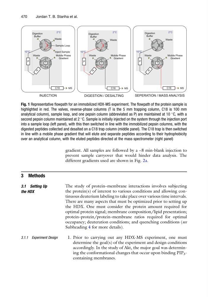
Fig. 1 Representative flowpath for an immobilized HDX-MS experiment. The flowpath of the protein sample is
highlighted in red. The valves, reverse-phase columns (T is the 5 mm trapping column, C18 is 100 mm
analytical column), sample loop, and one pepsin column (abbreviated as P) are maintained at 10
C, with a
second pepsin column maintained at 2
C. Sample is initially injected on the system through the injection port
into a sample loop (left panel), with this then switched in line with the immobilized pepsin columns, with the
digested peptides collected and desalted on a C18 trap column (middle panel). The C18 trap is then switched
in line with a mobile phase gradient that will elute and separate peptides according to their hydrophobicity
over an analytical column, with the eluted peptides directed at the mass spectrometer (right panel)
470
Jordan T. B. Stariha et al.
prevent sample carryover that would hinder data analysis. The
different gradients used are shown in Fig. 2a.
3 Methods
3.1 Setting Up
the HDX
The study of protein–membrane interactions involves subjecting
the protein(s) of interest to various conditions and allowing continuous deuterium labeling to take place over various time intervals.
There are many aspects that must be optimized prior to setting up
the HDX. One must consider the protein amount required for
optimal protein signal; membrane composition/lipid presentation;
protein–protein/protein–membrane ratios required for optimal
occupancy; deuteration conditions; and quenching conditions (see
Subheading 4 for more details).
3.1.1 Experiment Design
1. Prior to carrying out any HDX-MS experiment, one must
determine the goal(s) of the experiment and design conditions
accordingly. In the study of Akt, the major goal was determining the conformational changes that occur upon binding PIP 3 -
containing membranes.
INJECTION
Inject Sample
Digestion
Buffer
1
2
3
4
5
6
1
2
3
4
5
6 T
Waste
Sample Loop
P
P
Mobile Phase
Gradient
Waste
MS
DIGESTION / DESALTING
1
2
3
4
5
6
1
2
3
4
5
6 T
Waste
P
Mobile Phase
Gradient
Waste
MS
SEPERATION / MASS ANALYSIS
1
2
3
4
5
6
1
2
3
4
5
6 T
Waste
P
P
Mobile Phase
Gradient
Waste
MS
C18
C18
C18
2°C
10°C
10°C
P
10°C
Digestion
Buffer
Digestion
Buffer
2°C
2°C
Fig. 1 Representative flowpath for an immobilized HDX-MS experiment. The flowpath of the protein sample is
highlighted in red. The valves, reverse-phase columns (T is the 5 mm trapping column, C18 is 100 mm
analytical column), sample loop, and one pepsin column (abbreviated as P) are maintained at 10
C, with a
second pepsin column maintained at 2
C. Sample is initially injected on the system through the injection port
into a sample loop (left panel), with this then switched in line with the immobilized pepsin columns, with the
digested peptides collected and desalted on a C18 trap column (middle panel). The C18 trap is then switched
in line with a mobile phase gradient that will elute and separate peptides according to their hydrophobicity
over an analytical column, with the eluted peptides directed at the mass spectrometer (right panel)
470
Jordan T. B. Stariha et al.