

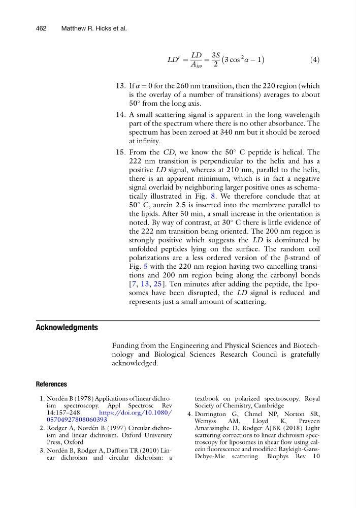
LD
r
¼
LD
A iso
¼
3S
2
3 cos
2
α À 1
À
Á
ð4Þ
13. If α ¼ 0 for the 260 nm transition, then the 220 region (which
is the overlay of a number of transitions) averages to about
50
from the long axis.
14. A small scattering signal is apparent in the long wavelength
part of the spectrum where there is no other absorbance. The
spectrum has been zeroed at 340 nm but it should be zeroed
at infinity.
15. From the CD, we know the 50
C peptide is helical. The
222 nm transition is perpendicular to the helix and has a
positive LD signal, whereas at 210 nm, parallel to the helix,
there is an apparent minimum, which is in fact a negative
signal overlaid by neighboring larger positive ones as schematically illustrated in Fig. 8. We therefore conclude that at
50
C, aurein 2.5 is inserted into the membrane parallel to
the lipids. After 50 min, a small increase in the orientation is
noted. By way of contrast, at 30
C there is little evidence of
the 222 nm transition being oriented. The 200 nm region is
strongly positive which suggests the LD is dominated by
unfolded peptides lying on the surface. The random coil
polarizations are a less ordered version of the β-strand of
Fig. 5 with the 220 nm region having two cancelling transitions and 200 nm region being along the carbonyl bonds
[7, 13, 25]. Ten minutes after adding the peptide, the liposomes have been disrupted, the LD signal is reduced and
represents just a small amount of scattering.
Acknowledgments
Funding from the Engineering and Physical Sciences and Biotechnology and Biological Sciences Research Council is gratefully
acknowledged.
References
1. Norde ´n B (1978) Applications of linear dichroism spectroscopy. Appl Spectrosc Rev
14:157–248.
https://doi.org/10.1080/
05704927808060393
2. Rodger A, Norde ´n B (1997) Circular dichroism and linear dichroism. Oxford University
Press, Oxford
3. Norde ´n B, Rodger A, Dafforn TR (2010) Linear dichroism and circular dichroism: a
textbook on polarized spectroscopy. Royal
Society of Chemistry, Cambridge
4. Dorrington G, Chmel NP, Norton SR,
Wemyss
AM,
Lloyd
K,
Praveen
Amarasinghe D, Rodger AJBR (2018) Light
scattering corrections to linear dichroism spectroscopy for liposomes in shear flow using calcein fluorescence and modified Rayleigh-GansDebye-Mie scattering. Biophys Rev 10
462
Matthew R. Hicks et al.
r
¼
LD
A iso
¼
3S
2
3 cos
2
α À 1
À
Á
ð4Þ
13. If α ¼ 0 for the 260 nm transition, then the 220 region (which
is the overlay of a number of transitions) averages to about
50
from the long axis.
14. A small scattering signal is apparent in the long wavelength
part of the spectrum where there is no other absorbance. The
spectrum has been zeroed at 340 nm but it should be zeroed
at infinity.
15. From the CD, we know the 50
C peptide is helical. The
222 nm transition is perpendicular to the helix and has a
positive LD signal, whereas at 210 nm, parallel to the helix,
there is an apparent minimum, which is in fact a negative
signal overlaid by neighboring larger positive ones as schematically illustrated in Fig. 8. We therefore conclude that at
50
C, aurein 2.5 is inserted into the membrane parallel to
the lipids. After 50 min, a small increase in the orientation is
noted. By way of contrast, at 30
C there is little evidence of
the 222 nm transition being oriented. The 200 nm region is
strongly positive which suggests the LD is dominated by
unfolded peptides lying on the surface. The random coil
polarizations are a less ordered version of the β-strand of
Fig. 5 with the 220 nm region having two cancelling transitions and 200 nm region being along the carbonyl bonds
[7, 13, 25]. Ten minutes after adding the peptide, the liposomes have been disrupted, the LD signal is reduced and
represents just a small amount of scattering.
Acknowledgments
Funding from the Engineering and Physical Sciences and Biotechnology and Biological Sciences Research Council is gratefully
acknowledged.
References
1. Norde ´n B (1978) Applications of linear dichroism spectroscopy. Appl Spectrosc Rev
14:157–248.
https://doi.org/10.1080/
05704927808060393
2. Rodger A, Norde ´n B (1997) Circular dichroism and linear dichroism. Oxford University
Press, Oxford
3. Norde ´n B, Rodger A, Dafforn TR (2010) Linear dichroism and circular dichroism: a
textbook on polarized spectroscopy. Royal
Society of Chemistry, Cambridge
4. Dorrington G, Chmel NP, Norton SR,
Wemyss
AM,
Lloyd
K,
Praveen
Amarasinghe D, Rodger AJBR (2018) Light
scattering corrections to linear dichroism spectroscopy for liposomes in shear flow using calcein fluorescence and modified Rayleigh-GansDebye-Mie scattering. Biophys Rev 10
462
Matthew R. Hicks et al.