

2. It is sometimes convenient to add a peptide or small molecule
analyte to the starting chloroform solution as this facilitates its
incorporation in the membrane.
3. It should be noted that Cl
À absorbs light below about 215 nm,
so concentrations higher than 10 mM should be avoided if
possible. Although Cl
À and other ions do not have an intrinsic
LD signal, they can mask the LD signal if they absorb too
much. Methods to remove Cl
À include dialysis, filtration with
appropriate membranes and columns. F
À can often be used to
substitute for the ionic strength effect of Cl
À .
4. Dynamic light scattering is a good method to check your
liposome size. It generally proves to be slightly higher than
the pore size.
5. Approximately 70–80 μL sample volume is usually sufficient in
a micro-volume capillary LD cell. It is possible to reduce the
volume, but ensure the light beam is not incident on the
meniscus of the sample as this introduces light scattering and
other artifacts.
6. Note that different lipids and analytes have different insertion
kinetics (taking from <1 s to overnight to insert). Also some
peptides destroy the liposomes (a mode of action of some
antimicrobial peptides). So consideration should be given
to when to collect spectroscopic data.
180
200
LD
220
240
260
50 min
10 min
5 min
Aurein 2.5 in DPPC/DPPG 30°
Aurein 2.5 in DPPC/DPPG 50°
Wavelength/nm
280
300
320
340
0
0.0005
0.001
0.0015
0.002
0.0025
0.003
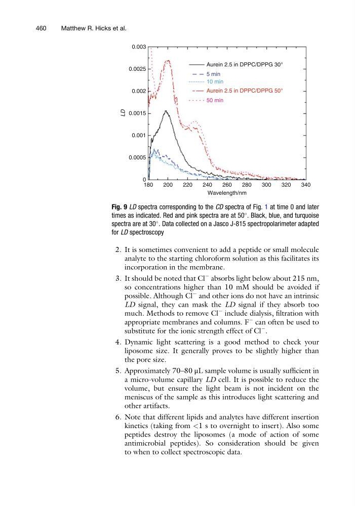
Fig. 9 LD spectra corresponding to the CD spectra of Fig. 1 at time 0 and later
times as indicated. Red and pink spectra are at 50
. Black, blue, and turquoise
spectra are at 30
. Data collected on a Jasco J-815 spectropolarimeter adapted
for LD spectroscopy
460
Matthew R. Hicks et al.
analyte to the starting chloroform solution as this facilitates its
incorporation in the membrane.
3. It should be noted that Cl
À absorbs light below about 215 nm,
so concentrations higher than 10 mM should be avoided if
possible. Although Cl
À and other ions do not have an intrinsic
LD signal, they can mask the LD signal if they absorb too
much. Methods to remove Cl
À include dialysis, filtration with
appropriate membranes and columns. F
À can often be used to
substitute for the ionic strength effect of Cl
À .
4. Dynamic light scattering is a good method to check your
liposome size. It generally proves to be slightly higher than
the pore size.
5. Approximately 70–80 μL sample volume is usually sufficient in
a micro-volume capillary LD cell. It is possible to reduce the
volume, but ensure the light beam is not incident on the
meniscus of the sample as this introduces light scattering and
other artifacts.
6. Note that different lipids and analytes have different insertion
kinetics (taking from <1 s to overnight to insert). Also some
peptides destroy the liposomes (a mode of action of some
antimicrobial peptides). So consideration should be given
to when to collect spectroscopic data.
180
200
LD
220
240
260
50 min
10 min
5 min
Aurein 2.5 in DPPC/DPPG 30°
Aurein 2.5 in DPPC/DPPG 50°
Wavelength/nm
280
300
320
340
0
0.0005
0.001
0.0015
0.002
0.0025
0.003
Fig. 9 LD spectra corresponding to the CD spectra of Fig. 1 at time 0 and later
times as indicated. Red and pink spectra are at 50
. Black, blue, and turquoise
spectra are at 30
. Data collected on a Jasco J-815 spectropolarimeter adapted
for LD spectroscopy
460
Matthew R. Hicks et al.