

and LAMBDA™ 465 (Perkin Elmer) offer simple procedures to
measure absorbance and accurately determine RNA concentration
and purity. In cases where the RNA oligonucleotide contains a
modified base that absorbs at 340 nm, baseline correction should
be turned off on the NanoDrop™ to obtain more accurate results.
3.6 Construct Design
Many RNA–protein complexes comprise multiple domains
connected via flexible peptide linkers. To improve the chances of
successfully crystallizing a novel RNA–protein complex, it is important to produce and screen multiple construct variants with carefully selected protein domain boundaries, e.g., constructs for the
full-length protein or individual RBD, and to sample a variety of
different lengths of RNA [15]. Each construct should retain the key
features of the RNA–protein complex including the regions implicated in RNA–protein interactions and other functionality. Ideally,
the ability of constructs to bind RNA should be confirmed using
orthogonal biochemical and biophysical assays, e.g., the electrophoretic mobility shift assay (EMSA), surface plasmon resonance
(SPR), and nuclear magnetic resonance (NMR) (see Subheading
3.8). A BLAST search (https://blast.ncbi.nlm.nih.gov) can be used
to determine protein domain boundaries by comparing a protein
sequence with structurally characterized proteins and protein
domains in the PDB. In addition, limited proteolysis followed by
mass spectrometry analysis can be used to digest free and
RNA-bound protein in order to experimentally identify
RNA-binding proteolytic fragments. Subsequently, a series of constructs based on the experimentally determined domain boundaries
can be generated and evaluated for expression and solubility.
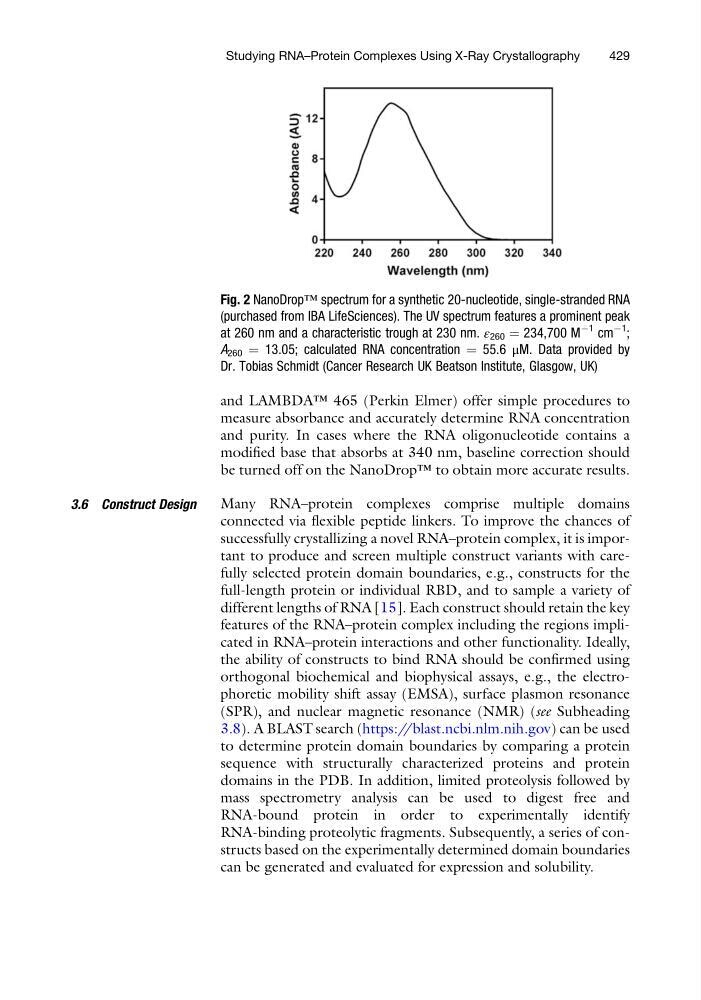
Fig. 2 NanoDrop™ spectrum for a synthetic 20-nucleotide, single-stranded RNA
(purchased from IBA LifeSciences). The UV spectrum features a prominent peak
at 260 nm and a characteristic trough at 230 nm. ε 260 ¼ 234,700 M
À1 cm
À1
;
A 260 ¼ 13.05; calculated RNA concentration ¼ 55.6 μM. Data provided by
Dr. Tobias Schmidt (Cancer Research UK Beatson Institute, Glasgow, UK)
Studying RNA–Protein Complexes Using X-Ray Crystallography
429
measure absorbance and accurately determine RNA concentration
and purity. In cases where the RNA oligonucleotide contains a
modified base that absorbs at 340 nm, baseline correction should
be turned off on the NanoDrop™ to obtain more accurate results.
3.6 Construct Design
Many RNA–protein complexes comprise multiple domains
connected via flexible peptide linkers. To improve the chances of
successfully crystallizing a novel RNA–protein complex, it is important to produce and screen multiple construct variants with carefully selected protein domain boundaries, e.g., constructs for the
full-length protein or individual RBD, and to sample a variety of
different lengths of RNA [15]. Each construct should retain the key
features of the RNA–protein complex including the regions implicated in RNA–protein interactions and other functionality. Ideally,
the ability of constructs to bind RNA should be confirmed using
orthogonal biochemical and biophysical assays, e.g., the electrophoretic mobility shift assay (EMSA), surface plasmon resonance
(SPR), and nuclear magnetic resonance (NMR) (see Subheading
3.8). A BLAST search (https://blast.ncbi.nlm.nih.gov) can be used
to determine protein domain boundaries by comparing a protein
sequence with structurally characterized proteins and protein
domains in the PDB. In addition, limited proteolysis followed by
mass spectrometry analysis can be used to digest free and
RNA-bound protein in order to experimentally identify
RNA-binding proteolytic fragments. Subsequently, a series of constructs based on the experimentally determined domain boundaries
can be generated and evaluated for expression and solubility.
Fig. 2 NanoDrop™ spectrum for a synthetic 20-nucleotide, single-stranded RNA
(purchased from IBA LifeSciences). The UV spectrum features a prominent peak
at 260 nm and a characteristic trough at 230 nm. ε 260 ¼ 234,700 M
À1 cm
À1
;
A 260 ¼ 13.05; calculated RNA concentration ¼ 55.6 μM. Data provided by
Dr. Tobias Schmidt (Cancer Research UK Beatson Institute, Glasgow, UK)
Studying RNA–Protein Complexes Using X-Ray Crystallography
429