

Aromatic side chains and disulfide bonds are the chromophores
in the near-UV regime. As the signal arising from these groups is
very sensitive to their environment, information on the overall
tertiary structure of the protein can be retrieved from these spectra
and even subtle changes in the structure can be detected, e.g., that
may occur upon binding to a ligand or changes in buffer composition, which do not alter secondary structure elements.
Overall, CD spectroscopy can be used to determine the folding
state of a protein to compare the structure (1) of proteins from
different sources/batches, (2) of different protein variants, and
(3) of proteins before and after changes in the production process,
as well as to study protein stability by both chemical and thermal
denaturation allowing the determination of thermodynamic parameters, to characterize conformational changes, and to analyze
protein:ligand interactions [18–20].
1.4 Assessing
Protein Stability
and Solubility
Generally, different types of “stability” can be distinguished. In the
QC context, it may be divided into “chemical” and “physical”
stability. Chemical stability describes how prone a protein is to
chemical modifications such as deamination and oxidation as well
as aspartic acid isomerization and nonreducible crosslinking; such
modifications can be checked by MS (see Subheading 1.2.3). In
order to facilitate reproducible downstream experimentation, it is
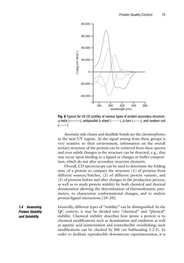
Fig. 6 Typical far-UV CD profiles of various types of protein secondary structure.
α-helix (––––––), antiparallel β-sheet (··········), β-turn (------), and random coil
(-·-·-·-·)
Protein Quality Control
15
in the near-UV regime. As the signal arising from these groups is
very sensitive to their environment, information on the overall
tertiary structure of the protein can be retrieved from these spectra
and even subtle changes in the structure can be detected, e.g., that
may occur upon binding to a ligand or changes in buffer composition, which do not alter secondary structure elements.
Overall, CD spectroscopy can be used to determine the folding
state of a protein to compare the structure (1) of proteins from
different sources/batches, (2) of different protein variants, and
(3) of proteins before and after changes in the production process,
as well as to study protein stability by both chemical and thermal
denaturation allowing the determination of thermodynamic parameters, to characterize conformational changes, and to analyze
protein:ligand interactions [18–20].
1.4 Assessing
Protein Stability
and Solubility
Generally, different types of “stability” can be distinguished. In the
QC context, it may be divided into “chemical” and “physical”
stability. Chemical stability describes how prone a protein is to
chemical modifications such as deamination and oxidation as well
as aspartic acid isomerization and nonreducible crosslinking; such
modifications can be checked by MS (see Subheading 1.2.3). In
order to facilitate reproducible downstream experimentation, it is
Fig. 6 Typical far-UV CD profiles of various types of protein secondary structure.
α-helix (––––––), antiparallel β-sheet (··········), β-turn (------), and random coil
(-·-·-·-·)
Protein Quality Control
15