

Knowledge of the expression and purification protocols and prior
storage conditions of the sample are very often also important in
interpreting experimental results and improving protein quality.
1.1 Preassessment
of Purity
and Concentration
Determination by
Ultraviolet
Spectroscopy
Proteins exhibit a characteristic ultraviolet (UV) absorption spectrum (Fig. 2) between 200 and 350 nm with most proteins having a
single maximum around 280 nm caused mainly by the aromatic
amino acids tyrosine (Tyr) and tryptophan (Trp). UV absorbance at
280 nm is routinely used to determine the protein concentration
via the Beer–Lambert law. However, this method will give inaccurate or erroneous results if the protein has no or only a few aromatic
amino acids or if it contains UV-absorbing nonprotein components
such as bound nucleic acids, nucleotide cofactors, porphyrins, or
iron–sulfur centers (see Note 1). In these cases, the absorbance at
205 or 214 nm, which is largely due to peptide bonds present in all
proteins, can be used instead to determine the protein
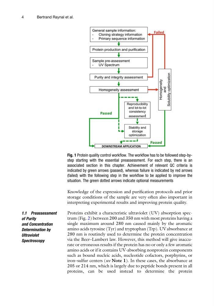
Fig. 1 Protein quality control workflow. The workflow has to be followed step-bystep starting with the essential preassessment. For each step, there is an
associated section in this chapter. Achievement of relevant QC criteria is
indicated by green arrows (passed), whereas failure is indicated by red arrows
(failed) with the following step in the workflow to be applied to improve the
situation. The green dotted arrows indicate optional measurements
4
Bertrand Raynal et al.
storage conditions of the sample are very often also important in
interpreting experimental results and improving protein quality.
1.1 Preassessment
of Purity
and Concentration
Determination by
Ultraviolet
Spectroscopy
Proteins exhibit a characteristic ultraviolet (UV) absorption spectrum (Fig. 2) between 200 and 350 nm with most proteins having a
single maximum around 280 nm caused mainly by the aromatic
amino acids tyrosine (Tyr) and tryptophan (Trp). UV absorbance at
280 nm is routinely used to determine the protein concentration
via the Beer–Lambert law. However, this method will give inaccurate or erroneous results if the protein has no or only a few aromatic
amino acids or if it contains UV-absorbing nonprotein components
such as bound nucleic acids, nucleotide cofactors, porphyrins, or
iron–sulfur centers (see Note 1). In these cases, the absorbance at
205 or 214 nm, which is largely due to peptide bonds present in all
proteins, can be used instead to determine the protein
Fig. 1 Protein quality control workflow. The workflow has to be followed step-bystep starting with the essential preassessment. For each step, there is an
associated section in this chapter. Achievement of relevant QC criteria is
indicated by green arrows (passed), whereas failure is indicated by red arrows
(failed) with the following step in the workflow to be applied to improve the
situation. The green dotted arrows indicate optional measurements
4
Bertrand Raynal et al.