

interactions [1]. One reason appears to be flexibility in binding,
both regarding structure (plasticity with regard to bound conformation) and number of binding partners (promiscuity). Because of
this, several biophysical studies of IDP interactions have tried to
address the mechanism(s) of binding and find quantitative evidence
for these predicted advantages of disorder [4].
IDPs and their interactions have been studied using the whole
battery of available biophysical techniques such as circular dichroism, calorimetry, fluorescence spectroscopy, single molecule techniques, and NMR. The latter is particularly powerful, since it
provides both structural and dynamic information [5], but a combination of multiple experimental approaches is always preferable
when trying to deduce mechanisms. Importantly, kinetics is the
only method that can unambiguously determine mechanisms in
protein–protein interactions, protein folding, and all other
biological reactions [6]. This chapter considers how to study interactions involving IDPs with the goal of determining mechanism, in
particular the collection and analysis of kinetic data obtained using
stopped-flow spectroscopy. We discuss basic principles of the methodology, experimental design, caveats and limitations of the
approach, and the analysis and interpretation of the data. While
we describe stopped flow, the approach is valid for other kinetic
methods.
1.2 A Kinetic
Approach to Assess
Mechanism
The association constant (K a ) between biomolecules tells us how
much they will bind to each other at equilibrium under a given set
of conditions and concentrations. Kinetic experiments tell us what
happens before this equilibrium is reached. If performed properly,
kinetic experiments can therefore provide clues to the reaction
mechanism; in the case of IDPs, what happens along the binding
trajectory as the free proteins interact, fold, and form a complex.
During protein folding and in binding reactions, it is surprisingly
common that intermediate states do not accumulate and therefore
cannot be detected easily in experiments. The process then consists
of one visible step, it is apparently “two state” (only unfolded and
folded, or free and bound, states are populated). Such a one-step
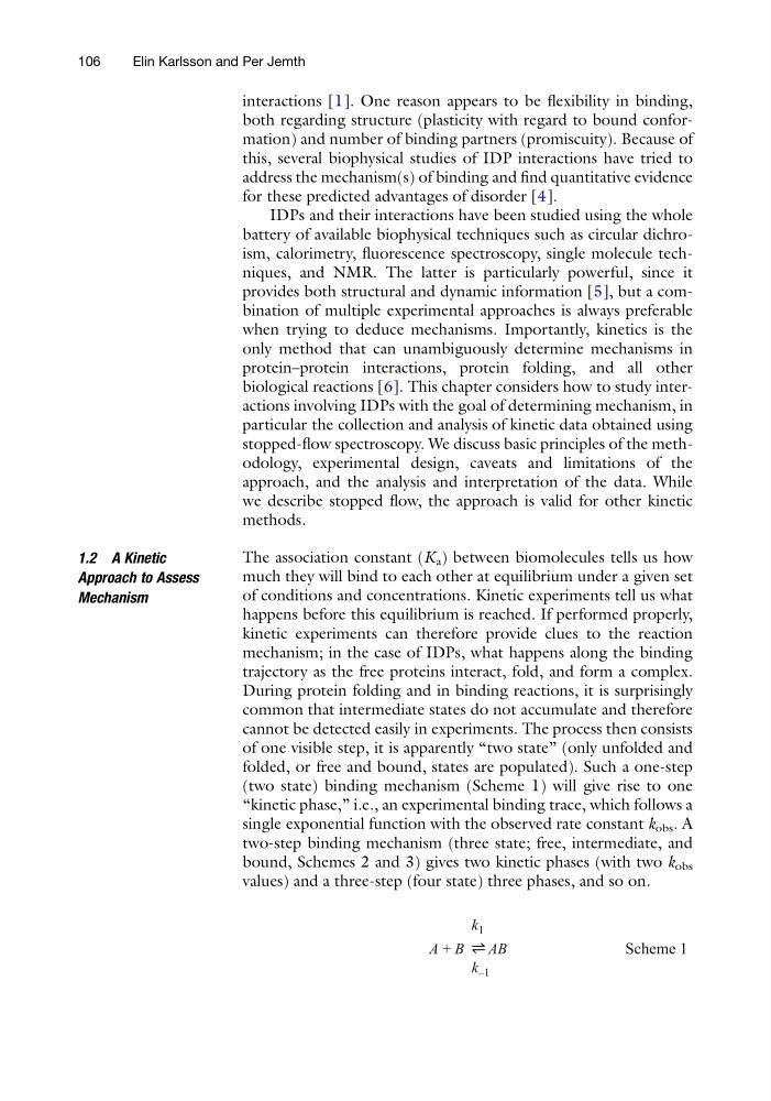
(two state) binding mechanism (Scheme 1) will give rise to one
“kinetic phase,” i.e., an experimental binding trace, which follows a
single exponential function with the observed rate constant k obs . A
two-step binding mechanism (three state; free, intermediate, and
bound, Schemes 2 and 3) gives two kinetic phases (with two k obs
values) and a three-step (four state) three phases, and so on.
Scheme 1
A + B
AB
k 1
k –1
106
Elin Karlsson and Per Jemth
both regarding structure (plasticity with regard to bound conformation) and number of binding partners (promiscuity). Because of
this, several biophysical studies of IDP interactions have tried to
address the mechanism(s) of binding and find quantitative evidence
for these predicted advantages of disorder [4].
IDPs and their interactions have been studied using the whole
battery of available biophysical techniques such as circular dichroism, calorimetry, fluorescence spectroscopy, single molecule techniques, and NMR. The latter is particularly powerful, since it
provides both structural and dynamic information [5], but a combination of multiple experimental approaches is always preferable
when trying to deduce mechanisms. Importantly, kinetics is the
only method that can unambiguously determine mechanisms in
protein–protein interactions, protein folding, and all other
biological reactions [6]. This chapter considers how to study interactions involving IDPs with the goal of determining mechanism, in
particular the collection and analysis of kinetic data obtained using
stopped-flow spectroscopy. We discuss basic principles of the methodology, experimental design, caveats and limitations of the
approach, and the analysis and interpretation of the data. While
we describe stopped flow, the approach is valid for other kinetic
methods.
1.2 A Kinetic
Approach to Assess
Mechanism
The association constant (K a ) between biomolecules tells us how
much they will bind to each other at equilibrium under a given set
of conditions and concentrations. Kinetic experiments tell us what
happens before this equilibrium is reached. If performed properly,
kinetic experiments can therefore provide clues to the reaction
mechanism; in the case of IDPs, what happens along the binding
trajectory as the free proteins interact, fold, and form a complex.
During protein folding and in binding reactions, it is surprisingly
common that intermediate states do not accumulate and therefore
cannot be detected easily in experiments. The process then consists
of one visible step, it is apparently “two state” (only unfolded and
folded, or free and bound, states are populated). Such a one-step
(two state) binding mechanism (Scheme 1) will give rise to one
“kinetic phase,” i.e., an experimental binding trace, which follows a
single exponential function with the observed rate constant k obs . A
two-step binding mechanism (three state; free, intermediate, and
bound, Schemes 2 and 3) gives two kinetic phases (with two k obs
values) and a three-step (four state) three phases, and so on.
Scheme 1
A + B
AB
k 1
k –1
106
Elin Karlsson and Per Jemth