

proteins such as calmodulin (P) can be studied by mixing with
an excess of a fluorescent Ca
2+ chelator (L) such as Quin
2, which forms a strongly fluorescing, high affinity 1:1 complex
with Ca
2+ [14].
12. Reactions such as those shown in Scheme C often exhibit
cooperativity in ligand binding. That is, for example, the affinity of X for P may be increased (positive cooperativity) or
decreased (negative cooperativity) when Y is also bound.
Changes in affinity may be caused by changes in either or
both of the rate constants defining the interaction with
X. Conservation of free energy for this scheme dictates that
(k À1 k À3 )/(k 1 k 3 ) must be equal to (k À2 k À4 )/(k 2 k 4 ).
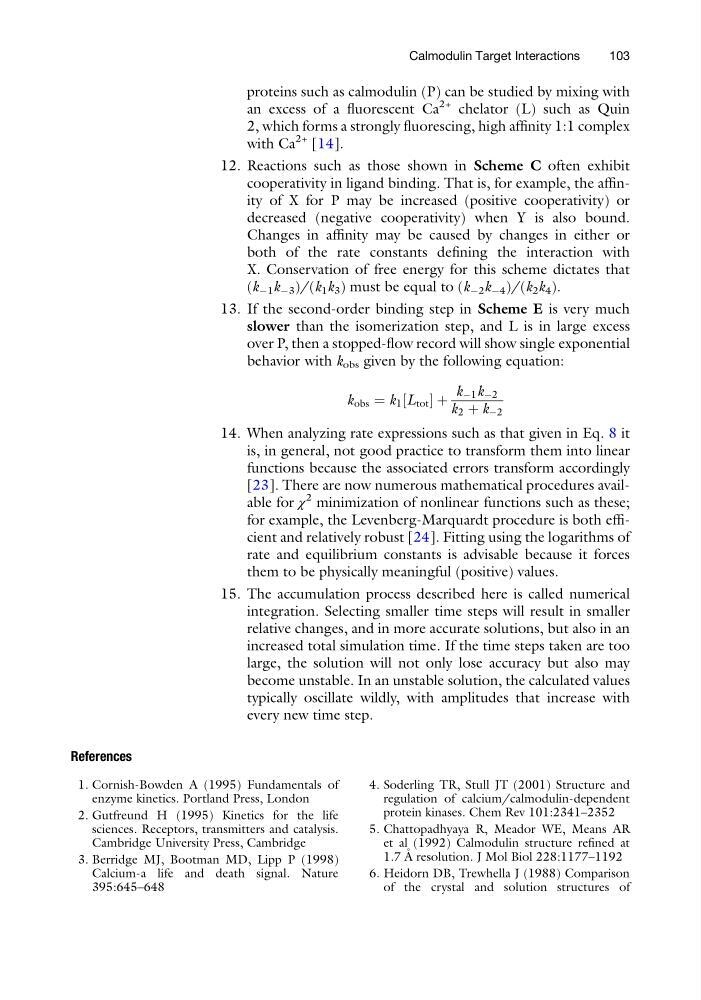
13. If the second-order binding step in Scheme E is very much
slower than the isomerization step, and L is in large excess
over P, then a stopped-flow record will show single exponential
behavior with k obs given by the following equation:
k obs ¼ k 1 L tot
½
þ
k À1 k À2
k 2 þ k À2
14. When analyzing rate expressions such as that given in Eq. 8 it
is, in general, not good practice to transform them into linear
functions because the associated errors transform accordingly
[23]. There are now numerous mathematical procedures available for χ
2 minimization of nonlinear functions such as these;
for example, the Levenberg-Marquardt procedure is both efficient and relatively robust [24]. Fitting using the logarithms of
rate and equilibrium constants is advisable because it forces
them to be physically meaningful (positive) values.
15. The accumulation process described here is called numerical
integration. Selecting smaller time steps will result in smaller
relative changes, and in more accurate solutions, but also in an
increased total simulation time. If the time steps taken are too
large, the solution will not only lose accuracy but also may
become unstable. In an unstable solution, the calculated values
typically oscillate wildly, with amplitudes that increase with
every new time step.
References
1. Cornish-Bowden A (1995) Fundamentals of
enzyme kinetics. Portland Press, London
2. Gutfreund H (1995) Kinetics for the life
sciences. Receptors, transmitters and catalysis.
Cambridge University Press, Cambridge
3. Berridge MJ, Bootman MD, Lipp P (1998)
Calcium-a life and death signal. Nature
395:645–648
4. Soderling TR, Stull JT (2001) Structure and
regulation of calcium/calmodulin-dependent
protein kinases. Chem Rev 101:2341–2352
5. Chattopadhyaya R, Meador WE, Means AR
et al (1992) Calmodulin structure refined at
1.7 A ˚ resolution. J Mol Biol 228:1177–1192
6. Heidorn DB, Trewhella J (1988) Comparison
of the crystal and solution structures of
Calmodulin Target Interactions
103
an excess of a fluorescent Ca
2+ chelator (L) such as Quin
2, which forms a strongly fluorescing, high affinity 1:1 complex
with Ca
2+ [14].
12. Reactions such as those shown in Scheme C often exhibit
cooperativity in ligand binding. That is, for example, the affinity of X for P may be increased (positive cooperativity) or
decreased (negative cooperativity) when Y is also bound.
Changes in affinity may be caused by changes in either or
both of the rate constants defining the interaction with
X. Conservation of free energy for this scheme dictates that
(k À1 k À3 )/(k 1 k 3 ) must be equal to (k À2 k À4 )/(k 2 k 4 ).
13. If the second-order binding step in Scheme E is very much
slower than the isomerization step, and L is in large excess
over P, then a stopped-flow record will show single exponential
behavior with k obs given by the following equation:
k obs ¼ k 1 L tot
½
þ
k À1 k À2
k 2 þ k À2
14. When analyzing rate expressions such as that given in Eq. 8 it
is, in general, not good practice to transform them into linear
functions because the associated errors transform accordingly
[23]. There are now numerous mathematical procedures available for χ
2 minimization of nonlinear functions such as these;
for example, the Levenberg-Marquardt procedure is both efficient and relatively robust [24]. Fitting using the logarithms of
rate and equilibrium constants is advisable because it forces
them to be physically meaningful (positive) values.
15. The accumulation process described here is called numerical
integration. Selecting smaller time steps will result in smaller
relative changes, and in more accurate solutions, but also in an
increased total simulation time. If the time steps taken are too
large, the solution will not only lose accuracy but also may
become unstable. In an unstable solution, the calculated values
typically oscillate wildly, with amplitudes that increase with
every new time step.
References
1. Cornish-Bowden A (1995) Fundamentals of
enzyme kinetics. Portland Press, London
2. Gutfreund H (1995) Kinetics for the life
sciences. Receptors, transmitters and catalysis.
Cambridge University Press, Cambridge
3. Berridge MJ, Bootman MD, Lipp P (1998)
Calcium-a life and death signal. Nature
395:645–648
4. Soderling TR, Stull JT (2001) Structure and
regulation of calcium/calmodulin-dependent
protein kinases. Chem Rev 101:2341–2352
5. Chattopadhyaya R, Meador WE, Means AR
et al (1992) Calmodulin structure refined at
1.7 A ˚ resolution. J Mol Biol 228:1177–1192
6. Heidorn DB, Trewhella J (1988) Comparison
of the crystal and solution structures of
Calmodulin Target Interactions
103