

221
case of spontaneous chromosomal deletion of the sbmA gene, the bacteria become
pyrrhocoricin resistant.
Hc-CATH is the first antimicrobial peptide cathelicidin, which was discovered in
sea snakes (Wei et al. 2015). Hc-CATH, as well as other peptides from the cathelicidins family, has a critical role against microbial infections in the vertebrates. The
structure of Hc-CATH is very stable and it is composed of 30 amino acids with two
α-helix regions (Wang et al. 2018). Hc-CATH demonstrates low cytotoxicity in
mammalian cells. Meanwhile, it shows high antibacterial activity in various Gram
negative and Gram positive bacteria such as Aeromonas salmonicida, Bacillus subtilis, Escherichia coli, Lactococcus garvieae, Klebsiella pneumoniae, Nocardia
asteroids, P. aeruginosa, Staphylococcus aureus, Shigella dysenteriae, Streptococcus
iniae and Vibrio (Vibrio vulnificus, Vibrio fluvialis, Vibrio splendidus).
LL-37 derives from human cathelicidin and it has an α-helical structure (Splith
and Neundorf 2011; Seil et al. 2010). It consists of 37 amino acid residues and it
demonstrates high antibacterial activity towards bacteria such as E. coli, L. monocytogenes, P. aeruginosa, S. aureus, and S. epidermidis. However, LL-37 also shows
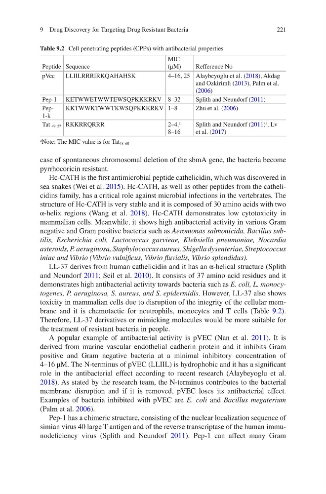
toxicity in mammalian cells due to disruption of the integrity of the cellular membrane and it is chemotactic for neutrophils, monocytes and T cells (Table 9.2).
Therefore, LL-37 derivatives or mimicking molecules would be more suitable for
the treatment of resistant bacteria in people.
A popular example of antibacterial activity is pVEC (Nan et al. 2011). It is
derived from murine vascular endothelial cadherin protein and it inhibits Gram
positive and Gram negative bacteria at a minimal inhibitory concentration of
4–16 μM. The N-terminus of pVEC (LLIIL) is hydrophobic and it has a significant
role in the antibacterial effect according to recent research (Alaybeyoglu et al.
2018). As stated by the research team, the N-terminus contributes to the bacterial
membrane disruption and if it is removed, pVEC loses its antibacterial effect.
Examples of bacteria inhibited with pVEC are E. coli and Bacillus megaterium
(Palm et al. 2006).
Pep-1 has a chimeric structure, consisting of the nuclear localization sequence of
simian virus 40 large T antigen and of the reverse transcriptase of the human immunodeficiency virus (Splith and Neundorf 2011). Pep-1 can affect many Gram
Table 9.2 Cell penetrating peptides (CPPs) with antibacterial properties
Peptide Sequence
MIC
(μM)
Refference No
pVec
LLIILRRRIRKQAHAHSK
4–16, 25 Alaybeyoglu et al. (2018), Akdag
and Ozkirimli (2013), Palm et al.
(2006)
Pep-1 KETWWETWWTEWSQPKKKRKV 8–32
Splith and Neundorf (2011)
Pep1- k
KKTWWKTWWTKWSQPKKKRKV 1–8
Zhu et al. (2006)
Tat 49–57 RKKRRQRRR
2–4,
a
8–16
Splith and Neundorf (2011)
a
, Lv
et al. (2017)
a Note: The MIC value is for Tat 48–60
9 Drug Discovery for Targeting Drug Resistant Bacteria
case of spontaneous chromosomal deletion of the sbmA gene, the bacteria become
pyrrhocoricin resistant.
Hc-CATH is the first antimicrobial peptide cathelicidin, which was discovered in
sea snakes (Wei et al. 2015). Hc-CATH, as well as other peptides from the cathelicidins family, has a critical role against microbial infections in the vertebrates. The
structure of Hc-CATH is very stable and it is composed of 30 amino acids with two
α-helix regions (Wang et al. 2018). Hc-CATH demonstrates low cytotoxicity in
mammalian cells. Meanwhile, it shows high antibacterial activity in various Gram
negative and Gram positive bacteria such as Aeromonas salmonicida, Bacillus subtilis, Escherichia coli, Lactococcus garvieae, Klebsiella pneumoniae, Nocardia
asteroids, P. aeruginosa, Staphylococcus aureus, Shigella dysenteriae, Streptococcus
iniae and Vibrio (Vibrio vulnificus, Vibrio fluvialis, Vibrio splendidus).
LL-37 derives from human cathelicidin and it has an α-helical structure (Splith
and Neundorf 2011; Seil et al. 2010). It consists of 37 amino acid residues and it
demonstrates high antibacterial activity towards bacteria such as E. coli, L. monocytogenes, P. aeruginosa, S. aureus, and S. epidermidis. However, LL-37 also shows
toxicity in mammalian cells due to disruption of the integrity of the cellular membrane and it is chemotactic for neutrophils, monocytes and T cells (Table 9.2).
Therefore, LL-37 derivatives or mimicking molecules would be more suitable for
the treatment of resistant bacteria in people.
A popular example of antibacterial activity is pVEC (Nan et al. 2011). It is
derived from murine vascular endothelial cadherin protein and it inhibits Gram
positive and Gram negative bacteria at a minimal inhibitory concentration of
4–16 μM. The N-terminus of pVEC (LLIIL) is hydrophobic and it has a significant
role in the antibacterial effect according to recent research (Alaybeyoglu et al.
2018). As stated by the research team, the N-terminus contributes to the bacterial
membrane disruption and if it is removed, pVEC loses its antibacterial effect.
Examples of bacteria inhibited with pVEC are E. coli and Bacillus megaterium
(Palm et al. 2006).
Pep-1 has a chimeric structure, consisting of the nuclear localization sequence of
simian virus 40 large T antigen and of the reverse transcriptase of the human immunodeficiency virus (Splith and Neundorf 2011). Pep-1 can affect many Gram
Table 9.2 Cell penetrating peptides (CPPs) with antibacterial properties
Peptide Sequence
MIC
(μM)
Refference No
pVec
LLIILRRRIRKQAHAHSK
4–16, 25 Alaybeyoglu et al. (2018), Akdag
and Ozkirimli (2013), Palm et al.
(2006)
Pep-1 KETWWETWWTEWSQPKKKRKV 8–32
Splith and Neundorf (2011)
Pep1- k
KKTWWKTWWTKWSQPKKKRKV 1–8
Zhu et al. (2006)
Tat 49–57 RKKRRQRRR
2–4,
a
8–16
Splith and Neundorf (2011)
a
, Lv
et al. (2017)
a Note: The MIC value is for Tat 48–60
9 Drug Discovery for Targeting Drug Resistant Bacteria