

113
dim and bright fluorescent cells and found that the dim cells were more tolerant to
ofloxacin as compared to the bright cells (Shah et al. 2006). This single fluorescent
protein based indicator enabled the identification of persister cells within the whole
population and isolation by fluorescence activated cell sorting (FACS). The destabilized green fluorescent protein can effectively differentiate the individual cells with
different growth rates when the expression of GFP des is under the control of rrnBP1
promoter. However, this indicator is unable to distinguish the persister cells and
dead cells if one wants to track the persister in different conditions, especially within
host cells. Combine rRNA-GFP des reporter with a constitutively expressed stable red
fluorescent protein (DsRed2) allowing for identifying the non growing cells and
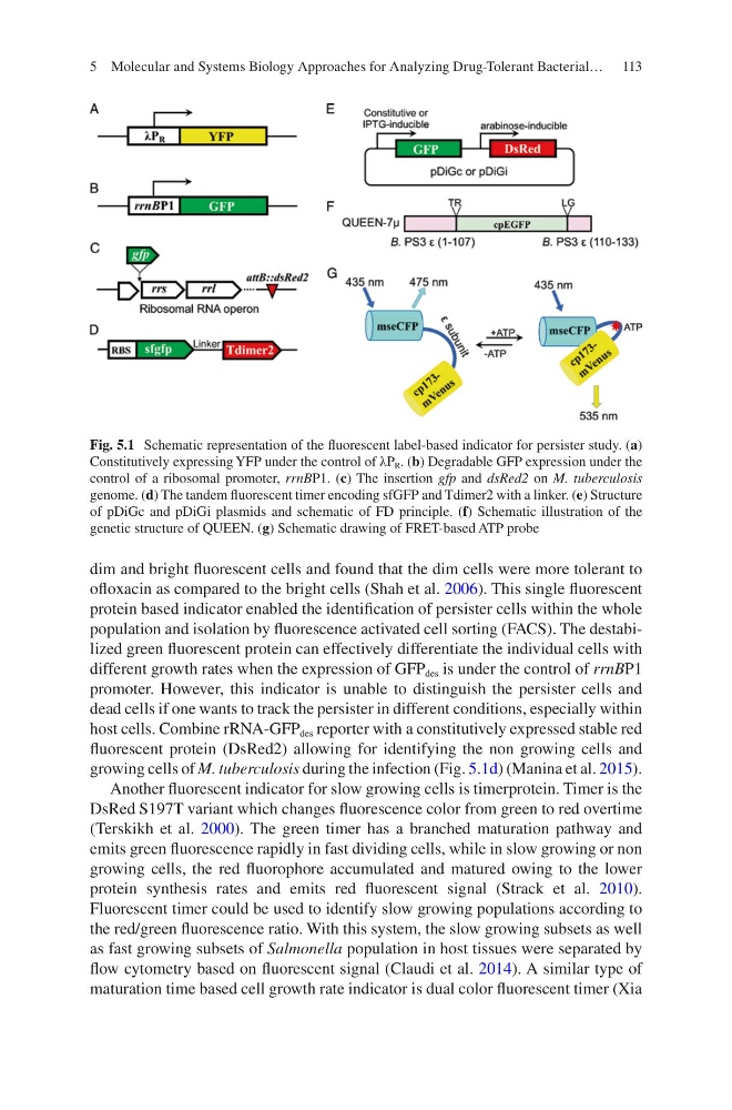
growing cells of M. tuberculosis during the infection (Fig. 5.1d) (Manina et al. 2015).
Another fluorescent indicator for slow growing cells is timerprotein. Timer is the
DsRed S197T variant which changes fluorescence color from green to red overtime
(Terskikh et al. 2000). The green timer has a branched maturation pathway and
emits green fluorescence rapidly in fast dividing cells, while in slow growing or non
growing cells, the red fluorophore accumulated and matured owing to the lower
protein synthesis rates and emits red fluorescent signal (Strack et al. 2010).
Fluorescent timer could be used to identify slow growing populations according to
the red/green fluorescence ratio. With this system, the slow growing subsets as well
as fast growing subsets of Salmonella population in host tissues were separated by
flow cytometry based on fluorescent signal (Claudi et al. 2014). A similar type of
maturation time based cell growth rate indicator is dual color fluorescent timer (Xia
Fig. 5.1 Schematic representation of the fluorescent label-based indicator for persister study. (a)
Constitutively expressing YFP under the control of λP R . (b) Degradable GFP expression under the
control of a ribosomal promoter, rrnBP1. (c) The insertion gfp and dsRed2 on M. tuberculosis
genome. (d) The tandem fluorescent timer encoding sfGFP and Tdimer2 with a linker. (e) Structure
of pDiGc and pDiGi plasmids and schematic of FD principle. (f) Schematic illustration of the
genetic structure of QUEEN. (g) Schematic drawing of FRET-based ATP probe
5 Molecular and Systems Biology Approaches for Analyzing Drug-Tolerant Bacterial…
dim and bright fluorescent cells and found that the dim cells were more tolerant to
ofloxacin as compared to the bright cells (Shah et al. 2006). This single fluorescent
protein based indicator enabled the identification of persister cells within the whole
population and isolation by fluorescence activated cell sorting (FACS). The destabilized green fluorescent protein can effectively differentiate the individual cells with
different growth rates when the expression of GFP des is under the control of rrnBP1
promoter. However, this indicator is unable to distinguish the persister cells and
dead cells if one wants to track the persister in different conditions, especially within
host cells. Combine rRNA-GFP des reporter with a constitutively expressed stable red
fluorescent protein (DsRed2) allowing for identifying the non growing cells and
growing cells of M. tuberculosis during the infection (Fig. 5.1d) (Manina et al. 2015).
Another fluorescent indicator for slow growing cells is timerprotein. Timer is the
DsRed S197T variant which changes fluorescence color from green to red overtime
(Terskikh et al. 2000). The green timer has a branched maturation pathway and
emits green fluorescence rapidly in fast dividing cells, while in slow growing or non
growing cells, the red fluorophore accumulated and matured owing to the lower
protein synthesis rates and emits red fluorescent signal (Strack et al. 2010).
Fluorescent timer could be used to identify slow growing populations according to
the red/green fluorescence ratio. With this system, the slow growing subsets as well
as fast growing subsets of Salmonella population in host tissues were separated by
flow cytometry based on fluorescent signal (Claudi et al. 2014). A similar type of
maturation time based cell growth rate indicator is dual color fluorescent timer (Xia
Fig. 5.1 Schematic representation of the fluorescent label-based indicator for persister study. (a)
Constitutively expressing YFP under the control of λP R . (b) Degradable GFP expression under the
control of a ribosomal promoter, rrnBP1. (c) The insertion gfp and dsRed2 on M. tuberculosis
genome. (d) The tandem fluorescent timer encoding sfGFP and Tdimer2 with a linker. (e) Structure
of pDiGc and pDiGi plasmids and schematic of FD principle. (f) Schematic illustration of the
genetic structure of QUEEN. (g) Schematic drawing of FRET-based ATP probe
5 Molecular and Systems Biology Approaches for Analyzing Drug-Tolerant Bacterial…