

112
5.3 Fluorescent Label Based Single Cell Approaches
Previous studies have focused on the persister cells in the whole population, which
only provide the measurement of averages and unable to analyse the specified subpopulation. To solve this problem, some studies start to observe the persister cells at
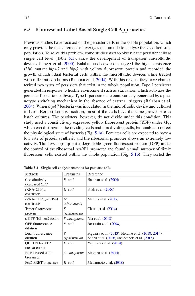
single cell level (Table 5.1), since the development of transparent microfluidic
devices (Unger et al. 2000). Balaban and coworkers tagged the high persistence
(hip) mutant hipA7 and hipQ with yellow fluorescent protein and recorded the
growth of individual bacterial cells within the microfluidic devices while treated
with different conditions (Balaban et al. 2004). With this device, they have characterized two types of persisters that exist in the whole population. Type I persisters
generated in response to hostile environment such as starvation, which activates the
persister formation pathway. Type II persisters are continuously generated by a phenotype switching mechanism in the absence of external triggers (Balaban et al.
2004). When hipA7 bacteria was inoculated in the microfluidic device and cultured
in Luria-Bertani Lennox medium, most of the cells have the same growth rate as
batch cultures. The persisters, however, do not divide under this condition. This
study used a constitutively expressed yellow fluorescent protein (YFP) under λP R ,
which can distinguish the dividing cells and non dividing cells, but unable to reflect
the physiological state of bacteria (Fig. 5.1a). Persister cells are expected to have a
low rate of protein synthesis and the ribosomal promoter shows an extremely low
activity. The Lewis group put a degradable green fluorescent protein (GFP) under
the control of the ribosomal rrnBP1 promoter and found a small number of dimly
fluorescent cells existed within the whole population (Fig. 5.1b). They sorted the
Table 5.1 Single cell analysis methods for persister cells
Methods
Organisms
Reference
Constitutively
expressed YFP
E. coli
Balaban et al. (2004)
rRNA-GFP des
constructs
E. coli
Shah et al. (2006)
rRNA-GFP des -DsRed
constructs
M.
tuberculosis
Manina et al. (2015)
Timer fluorescent
protein
S.
typhimurium
Claudi et al. (2014)
sfGFP-Tdimer2 fusion P. aeruginosa Xia et al. (2018)
GFP fluorescence
dilution
E. coli
Roostalu et al. (2008)
Dual fluorescence
dilution
S.
typhimurium
Figueira et al. (2013), Helaine et al. (2010, 2014),
Saliba et al. (2016) and Stapels et al. (2018)
QUEEN for ATP
measurement
E. coli
Yaginuma et al. (2014)
FRET-based ATP
biosensor
M. smegmatis Maglica et al. (2015)
FtsZ-FRET biosensor
E. coli
Matsumoto et al. (2018)
X. Duan et al.
5.3 Fluorescent Label Based Single Cell Approaches
Previous studies have focused on the persister cells in the whole population, which
only provide the measurement of averages and unable to analyse the specified subpopulation. To solve this problem, some studies start to observe the persister cells at
single cell level (Table 5.1), since the development of transparent microfluidic
devices (Unger et al. 2000). Balaban and coworkers tagged the high persistence
(hip) mutant hipA7 and hipQ with yellow fluorescent protein and recorded the
growth of individual bacterial cells within the microfluidic devices while treated
with different conditions (Balaban et al. 2004). With this device, they have characterized two types of persisters that exist in the whole population. Type I persisters
generated in response to hostile environment such as starvation, which activates the
persister formation pathway. Type II persisters are continuously generated by a phenotype switching mechanism in the absence of external triggers (Balaban et al.
2004). When hipA7 bacteria was inoculated in the microfluidic device and cultured
in Luria-Bertani Lennox medium, most of the cells have the same growth rate as
batch cultures. The persisters, however, do not divide under this condition. This
study used a constitutively expressed yellow fluorescent protein (YFP) under λP R ,
which can distinguish the dividing cells and non dividing cells, but unable to reflect
the physiological state of bacteria (Fig. 5.1a). Persister cells are expected to have a
low rate of protein synthesis and the ribosomal promoter shows an extremely low
activity. The Lewis group put a degradable green fluorescent protein (GFP) under
the control of the ribosomal rrnBP1 promoter and found a small number of dimly
fluorescent cells existed within the whole population (Fig. 5.1b). They sorted the
Table 5.1 Single cell analysis methods for persister cells
Methods
Organisms
Reference
Constitutively
expressed YFP
E. coli
Balaban et al. (2004)
rRNA-GFP des
constructs
E. coli
Shah et al. (2006)
rRNA-GFP des -DsRed
constructs
M.
tuberculosis
Manina et al. (2015)
Timer fluorescent
protein
S.
typhimurium
Claudi et al. (2014)
sfGFP-Tdimer2 fusion P. aeruginosa Xia et al. (2018)
GFP fluorescence
dilution
E. coli
Roostalu et al. (2008)
Dual fluorescence
dilution
S.
typhimurium
Figueira et al. (2013), Helaine et al. (2010, 2014),
Saliba et al. (2016) and Stapels et al. (2018)
QUEEN for ATP
measurement
E. coli
Yaginuma et al. (2014)
FRET-based ATP
biosensor
M. smegmatis Maglica et al. (2015)
FtsZ-FRET biosensor
E. coli
Matsumoto et al. (2018)
X. Duan et al.