

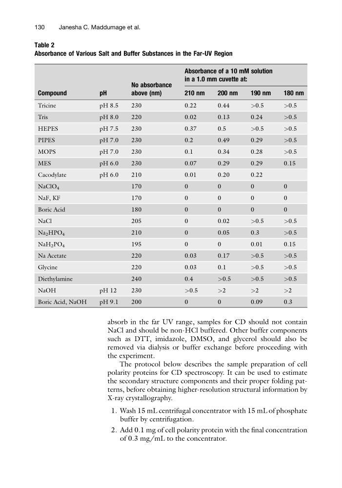
absorb in the far UV range, samples for CD should not contain
NaCl and should be non-HCl buffered. Other buffer components
such as DTT, imidazole, DMSO, and glycerol should also be
removed via dialysis or buffer exchange before proceeding with
the experiment.
The protocol below describes the sample preparation of cell
polarity proteins for CD spectroscopy. It can be used to estimate
the secondary structure components and their proper folding patterns, before obtaining higher-resolution structural information by
X-ray crystallography.
1. Wash 15 mL centrifugal concentrator with 15 mL of phosphate
buffer by centrifugation.
2. Add 0.1 mg of cell polarity protein with the final concentration
of 0.3 mg/mL to the concentrator.
Table 2
Absorbance of Various Salt and Buffer Substances in the Far-UV Region
Compound
pH
No absorbance
above (nm)
Absorbance of a 10 mM solution
in a 1.0 mm cuvette at:
210 nm
200 nm
190 nm
180 nm
Tricine
pH 8.5
230
0.22
0.44
>0.5
>0.5
Tris
pH 8.0
220
0.02
0.13
0.24
>0.5
HEPES
pH 7.5
230
0.37
0.5
>0.5
>0.5
PIPES
pH 7.0
230
0.2
0.49
0.29
>0.5
MOPS
pH 7.0
230
0.1
0.34
0.28
>0.5
MES
pH 6.0
230
0.07
0.29
0.29
0.15
Cacodylate
pH 6.0
210
0.01
0.20
0.22
NaClO 4
1 7 0
0
0
0
0
N a F ,K F
1 7 0
0
0
0
0
Boric Acid
180
0
0
0
0
NaCl
205
0
0.02
>0.5
>0.5
Na 2 HPO 4
210
0
0.05
0.3
>0.5
NaH 2 PO 4
195
0
0
0.01
0.15
Na Acetate
220
0.03
0.17
>0.5
>0.5
Glycine
220
0.03
0.1
>0.5
>0.5
Diethylamine
240
0.4
>0.5
>0.5
>0.5
NaOH
pH 12
230
>0.5
>2
>2
>2
Boric Acid, NaOH
pH 9.1
200
0
0
0.09
0.3
130
Janesha C. Maddumage et al.
NaCl and should be non-HCl buffered. Other buffer components
such as DTT, imidazole, DMSO, and glycerol should also be
removed via dialysis or buffer exchange before proceeding with
the experiment.
The protocol below describes the sample preparation of cell
polarity proteins for CD spectroscopy. It can be used to estimate
the secondary structure components and their proper folding patterns, before obtaining higher-resolution structural information by
X-ray crystallography.
1. Wash 15 mL centrifugal concentrator with 15 mL of phosphate
buffer by centrifugation.
2. Add 0.1 mg of cell polarity protein with the final concentration
of 0.3 mg/mL to the concentrator.
Table 2
Absorbance of Various Salt and Buffer Substances in the Far-UV Region
Compound
pH
No absorbance
above (nm)
Absorbance of a 10 mM solution
in a 1.0 mm cuvette at:
210 nm
200 nm
190 nm
180 nm
Tricine
pH 8.5
230
0.22
0.44
>0.5
>0.5
Tris
pH 8.0
220
0.02
0.13
0.24
>0.5
HEPES
pH 7.5
230
0.37
0.5
>0.5
>0.5
PIPES
pH 7.0
230
0.2
0.49
0.29
>0.5
MOPS
pH 7.0
230
0.1
0.34
0.28
>0.5
MES
pH 6.0
230
0.07
0.29
0.29
0.15
Cacodylate
pH 6.0
210
0.01
0.20
0.22
NaClO 4
1 7 0
0
0
0
0
N a F ,K F
1 7 0
0
0
0
0
Boric Acid
180
0
0
0
0
NaCl
205
0
0.02
>0.5
>0.5
Na 2 HPO 4
210
0
0.05
0.3
>0.5
NaH 2 PO 4
195
0
0
0.01
0.15
Na Acetate
220
0.03
0.17
>0.5
>0.5
Glycine
220
0.03
0.1
>0.5
>0.5
Diethylamine
240
0.4
>0.5
>0.5
>0.5
NaOH
pH 12
230
>0.5
>2
>2
>2
Boric Acid, NaOH
pH 9.1
200
0
0
0.09
0.3
130
Janesha C. Maddumage et al.