

3.16 Staining of Cell
Nucleus and Plasma
Membrane on Live/
Fixed Cells
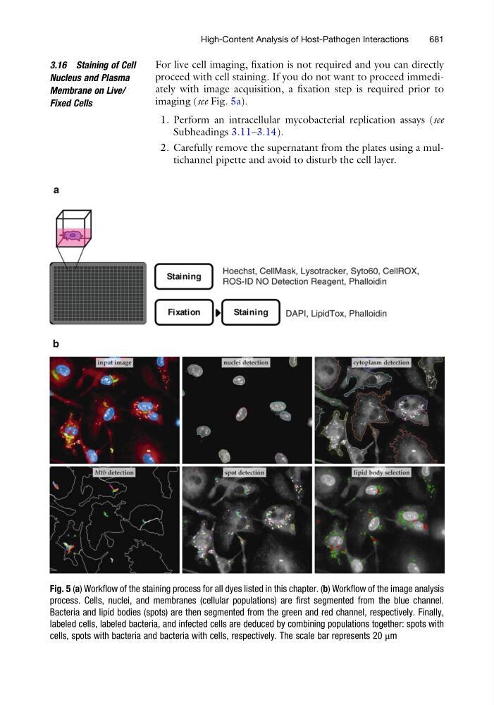
For live cell imaging, fixation is not required and you can directly
proceed with cell staining. If you do not want to proceed immediately with image acquisition, a fixation step is required prior to
imaging (see Fig. 5a).
1. Perform an intracellular mycobacterial replication assays (see
Subheadings 3.11–3.14).
2. Carefully remove the supernatant from the plates using a multichannel pipette and avoid to disturb the cell layer.
Fig. 5 (a) Workflow of the staining process for all dyes listed in this chapter. (b) Workflow of the image analysis
process. Cells, nuclei, and membranes (cellular populations) are first segmented from the blue channel.
Bacteria and lipid bodies (spots) are then segmented from the green and red channel, respectively. Finally,
labeled cells, labeled bacteria, and infected cells are deduced by combining populations together: spots with
cells, spots with bacteria and bacteria with cells, respectively. The scale bar represents 20 μm
High-Content Analysis of Host-Pathogen Interactions
681