

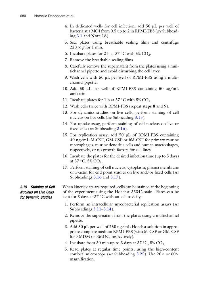
4. In dedicated wells for cell infection: add 50 μL per well of
bacteria at a MOI from 0.5 up to 2 in RPMI-FBS (see Subheading 3.1 and Note 18).
5. Seal plates using breathable sealing films and centrifuge
220 Â g for 1 min.
6. Incubate plates for 2 h at 37
C with 5% CO 2 .
7. Remove the breathable sealing films.
8. Carefully remove the supernatant from the plates using a multichannel pipette and avoid disturbing the cell layer.
9. Wash cells with 50 μL per well of RPMI-FBS using a multichannel pipette.
10. Add 50 μL per well of RPMI-FBS containing 50 μg/mL
amikacin.
11. Incubate plates for 1 h at 37
C with 5% CO 2 .
12. Wash cells twice with RPMI-FBS (repeat steps 8 and 9).
13. For dynamics studies on live cells, perform staining of cell
nucleus on live cells (see Subheading 3.15).
14. For uptake assay, perform staining of cell nucleus on live or
fixed cells (see Subheading 3.16).
15. For replication assay, add 50 μL of RPMI-FBS containing
40 ng/mL M-CSF, GM-CSF or hM-CSF for primary murine
macrophages, murine dendritic cells and human macrophages,
respectively, or no growth factors for cell lines.
16. Incubate the plates for the desired infection time (up to 5 days)
at 37
C, 5% CO 2 .
17. Perform staining of cell nucleus, cytoplasm, plasma membrane
or F-actin for end point studies on live and/or fixed cells (see
Subheadings 3.16 and 3.17).
3.15 Staining of Cell
Nucleus on Live Cells
for Dynamic Studies
When kinetic data are required, cells can be stained at the beginning
of the experiment using the Hoechst 33342 stain. Plates can be
kept for 3 days at 37
C without cell toxicity.
1. Perform an intracellular mycobacterial replication assays (see
Subheadings 3.11–3.14).
2. Remove the supernatant from the plates using a multichannel
pipette.
3. Add 50 μL per well of 250 ng/mL Hoechst solution in appropriate complete medium RPMI-FBS (with M-CSF or GM-CSF
for BMDM or BMDC, respectively).
4. Incubate from 30 min up to 3 days at 37
C, 5% CO 2 .
5. Read plates at regular time points, using the high-content
confocal microscope (see Subheading 3.25). Use 20Â or 60Â
magnification.
680
Nathalie Deboosere et al.