

2. It should be noted that the widely used M. tuberculosis H37Rv
strain has genomic differences (e.g., in the two component
system gene phoR) that result in a lack of some aspects of
environmental-induced transcriptional upregulation observed
with the Erdman and CDC1551 strains [34].
3. M. tuberculosis is very sensitive to pH, and calibration of the pH
meter using pH 4, 7, and 10 standards before preparation of
media is thus important. Fresh pH standards should be used,
particularly in the case of the pH 10 standard, as the pH of this
buffer is changed by exposure to carbon dioxide present in air.
We use calibration buffer pouches, with buffer solutions used
within 1 week of opening.
4. It is important that care be taken when adjusting pH of
M. tuberculosis media so that the desired pH is not overshot
(requiring more than the minimal base addition). Use of an
acid such as HCl to adjust pH back down is to be avoided, as
such further addition changes the ionic strength of the
medium.
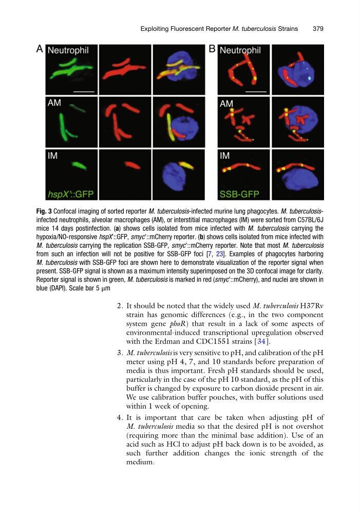
Fig. 3 Confocal imaging of sorted reporter M. tuberculosis-infected murine lung phagocytes. M. tuberculosisinfected neutrophils, alveolar macrophages (AM), or interstitial macrophages (IM) were sorted from C57BL/6J
mice 14 days postinfection. (a) shows cells isolated from mice infected with M. tuberculosis carrying the
hypoxia/NO-responsive hspX’::GFP, smyc’::mCherry reporter. (b) shows cells isolated from mice infected with
M. tuberculosis carrying the replication SSB-GFP, smyc’::mCherry reporter. Note that most M. tuberculosis
from such an infection will not be positive for SSB-GFP foci [7, 23]. Examples of phagocytes harboring
M. tuberculosis with SSB-GFP foci are shown here to demonstrate visualization of the reporter signal when
present. SSB-GFP signal is shown as a maximum intensity superimposed on the 3D confocal image for clarity.
Reporter signal is shown in green, M. tuberculosis is marked in red (smyc’::mCherry), and nuclei are shown in
blue (DAPI). Scale bar 5 μm
Exploiting Fluorescent Reporter M. tuberculosis Strains
379
strain has genomic differences (e.g., in the two component
system gene phoR) that result in a lack of some aspects of
environmental-induced transcriptional upregulation observed
with the Erdman and CDC1551 strains [34].
3. M. tuberculosis is very sensitive to pH, and calibration of the pH
meter using pH 4, 7, and 10 standards before preparation of
media is thus important. Fresh pH standards should be used,
particularly in the case of the pH 10 standard, as the pH of this
buffer is changed by exposure to carbon dioxide present in air.
We use calibration buffer pouches, with buffer solutions used
within 1 week of opening.
4. It is important that care be taken when adjusting pH of
M. tuberculosis media so that the desired pH is not overshot
(requiring more than the minimal base addition). Use of an
acid such as HCl to adjust pH back down is to be avoided, as
such further addition changes the ionic strength of the
medium.
Fig. 3 Confocal imaging of sorted reporter M. tuberculosis-infected murine lung phagocytes. M. tuberculosisinfected neutrophils, alveolar macrophages (AM), or interstitial macrophages (IM) were sorted from C57BL/6J
mice 14 days postinfection. (a) shows cells isolated from mice infected with M. tuberculosis carrying the
hypoxia/NO-responsive hspX’::GFP, smyc’::mCherry reporter. (b) shows cells isolated from mice infected with
M. tuberculosis carrying the replication SSB-GFP, smyc’::mCherry reporter. Note that most M. tuberculosis
from such an infection will not be positive for SSB-GFP foci [7, 23]. Examples of phagocytes harboring
M. tuberculosis with SSB-GFP foci are shown here to demonstrate visualization of the reporter signal when
present. SSB-GFP signal is shown as a maximum intensity superimposed on the 3D confocal image for clarity.
Reporter signal is shown in green, M. tuberculosis is marked in red (smyc’::mCherry), and nuclei are shown in
blue (DAPI). Scale bar 5 μm
Exploiting Fluorescent Reporter M. tuberculosis Strains
379