

16. Incubate on a nutator for 30 min at room temperature, protected from light.
17. Pellet cells at 3000 rpm (850 Â g) for 5 min.
18. Wash cell pellet once with 1 mL PBS, before resuspending in
100 μL PBS.
19. Transfer the cell suspensions to an 8-chambered cover glass
coated with poly-L-lysine (100 μg/mL), and leave for at least
1 h at room temperature to allow the cells to settle and attach
to the cover glass surface.
20. Visualize and quantify reporter M. tuberculosis signal as
described (see Subheading 3.5, steps 16–21), quantifying at
least 100 bacteria for each host cell type (Fig. 3).
4 Notes
1. This is only needed if the TOPO cloning steps in Subheadings
3.1 and 3.2 are utilized. While not essential, TOPO cloning
enables easy sequencing verification, long-term maintenance of
the verified construct, and increased efficiency at the ligation
step of cloning.
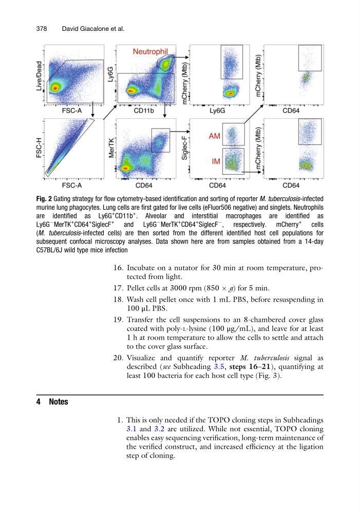
Fig. 2 Gating strategy for flow cytometry-based identification and sorting of reporter M. tuberculosis-infected
murine lung phagocytes. Lung cells are first gated for live cells (eFluor506 negative) and singlets. Neutrophils
are identified as Ly6G
+
CD11b
+
. Alveolar and interstitial macrophages are identified as
Ly6G
À
MerTK
+
CD64
+
SiglecF
+
and Ly6G
À
MerTK
+
CD64
+
SiglecF
À
, respectively. mCherry
+
cells
(M. tuberculosis-infected cells) are then sorted from the different identified host cell populations for
subsequent confocal microscopy analyses. Data shown here are from samples obtained from a 14-day
C57BL/6J wild type mice infection
378
David Giacalone et al.
17. Pellet cells at 3000 rpm (850 Â g) for 5 min.
18. Wash cell pellet once with 1 mL PBS, before resuspending in
100 μL PBS.
19. Transfer the cell suspensions to an 8-chambered cover glass
coated with poly-L-lysine (100 μg/mL), and leave for at least
1 h at room temperature to allow the cells to settle and attach
to the cover glass surface.
20. Visualize and quantify reporter M. tuberculosis signal as
described (see Subheading 3.5, steps 16–21), quantifying at
least 100 bacteria for each host cell type (Fig. 3).
4 Notes
1. This is only needed if the TOPO cloning steps in Subheadings
3.1 and 3.2 are utilized. While not essential, TOPO cloning
enables easy sequencing verification, long-term maintenance of
the verified construct, and increased efficiency at the ligation
step of cloning.
Fig. 2 Gating strategy for flow cytometry-based identification and sorting of reporter M. tuberculosis-infected
murine lung phagocytes. Lung cells are first gated for live cells (eFluor506 negative) and singlets. Neutrophils
are identified as Ly6G
+
CD11b
+
. Alveolar and interstitial macrophages are identified as
Ly6G
À
MerTK
+
CD64
+
SiglecF
+
and Ly6G
À
MerTK
+
CD64
+
SiglecF
À
, respectively. mCherry
+
cells
(M. tuberculosis-infected cells) are then sorted from the different identified host cell populations for
subsequent confocal microscopy analyses. Data shown here are from samples obtained from a 14-day
C57BL/6J wild type mice infection
378
David Giacalone et al.