

12. Allow the plates to dry, wrap them with 2 sheets of aluminum
foil and incubate the plates at 37
C for 21 days.
13. Successful integration of the plasmid into the oligo-directed
attP site will be evident if many more colonies appear on the
plates spread with cells that were electroporated with both
oligo and plasmid, relative to cells electroporated with just
the plasmid.
3.5 Verification
of ORBIT-Mediated
Recombinants
in M. tuberculosis
1. Pick four to six colonies with a sterile plastic loop and inoculate
each into 30 mL inkwell bottles containing 5 mL of
7H9-OADC-Tween 80 and 50 μg/mL hygromycin
(or 25 μg/mL zeocin).
2. After the OD 600 reaches ~1.0–1.5 (but no more), transfer
0.3 mL of the cultures to screw-capped tubes and heat inactivate the cells by total immersion of the tubes in hot metal bead
bath (85
C) for 50 min.
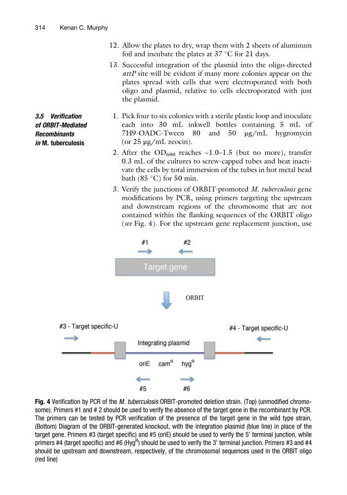
3. Verify the junctions of ORBIT-promoted M. tuberculosis gene
modifications by PCR, using primers targeting the upstream
and downstream regions of the chromosome that are not
contained within the flanking sequences of the ORBIT oligo
(see Fig. 4). For the upstream gene replacement junction, use
Fig. 4 Verification by PCR of the M. tuberculosis ORBIT-promoted deletion strain. (Top) (unmodified chromosome). Primers #1 and # 2 should be used to verify the absence of the target gene in the recombinant by PCR.
The primers can be tested by PCR verification of the presence of the target gene in the wild type strain.
(Bottom) Diagram of the ORBIT-generated knockout, with the integration plasmid (blue line) in place of the
target gene. Primers #3 (target specific) and #5 (oriE) should be used to verify the 5
0
terminal junction, while
primers #4 (target specific) and #6 (Hyg
R
) should be used to verify the 3
0
terminal junction. Primers #3 and #4
should be upstream and downstream, respectively, of the chromosomal sequences used in the ORBIT oligo
(red line)
314
Kenan C. Murphy
foil and incubate the plates at 37
C for 21 days.
13. Successful integration of the plasmid into the oligo-directed
attP site will be evident if many more colonies appear on the
plates spread with cells that were electroporated with both
oligo and plasmid, relative to cells electroporated with just
the plasmid.
3.5 Verification
of ORBIT-Mediated
Recombinants
in M. tuberculosis
1. Pick four to six colonies with a sterile plastic loop and inoculate
each into 30 mL inkwell bottles containing 5 mL of
7H9-OADC-Tween 80 and 50 μg/mL hygromycin
(or 25 μg/mL zeocin).
2. After the OD 600 reaches ~1.0–1.5 (but no more), transfer
0.3 mL of the cultures to screw-capped tubes and heat inactivate the cells by total immersion of the tubes in hot metal bead
bath (85
C) for 50 min.
3. Verify the junctions of ORBIT-promoted M. tuberculosis gene
modifications by PCR, using primers targeting the upstream
and downstream regions of the chromosome that are not
contained within the flanking sequences of the ORBIT oligo
(see Fig. 4). For the upstream gene replacement junction, use
Fig. 4 Verification by PCR of the M. tuberculosis ORBIT-promoted deletion strain. (Top) (unmodified chromosome). Primers #1 and # 2 should be used to verify the absence of the target gene in the recombinant by PCR.
The primers can be tested by PCR verification of the presence of the target gene in the wild type strain.
(Bottom) Diagram of the ORBIT-generated knockout, with the integration plasmid (blue line) in place of the
target gene. Primers #3 (target specific) and #5 (oriE) should be used to verify the 5
0
terminal junction, while
primers #4 (target specific) and #6 (Hyg
R
) should be used to verify the 3
0
terminal junction. Primers #3 and #4
should be upstream and downstream, respectively, of the chromosomal sequences used in the ORBIT oligo
(red line)
314
Kenan C. Murphy