

2.6 Soft Lithography
and Microfabrication
1. Silicon wafer mold (Fig. 1b).
2. Precision wafer handling tweezers.
3. Fast-drying glue.
4. Compressed air blow gun.
5. Plastic goblet 20 cl.
6. Disposable L-shaped spreader.
7. Polydimethylsiloxane (PDMS), heat-curable silicone elastomer
(Sylgard 184, Dow Corning).
8. Square petri dish, 120 Â 120 Â 17 mm.
9. Vacuum chamber.
10. Baking oven.
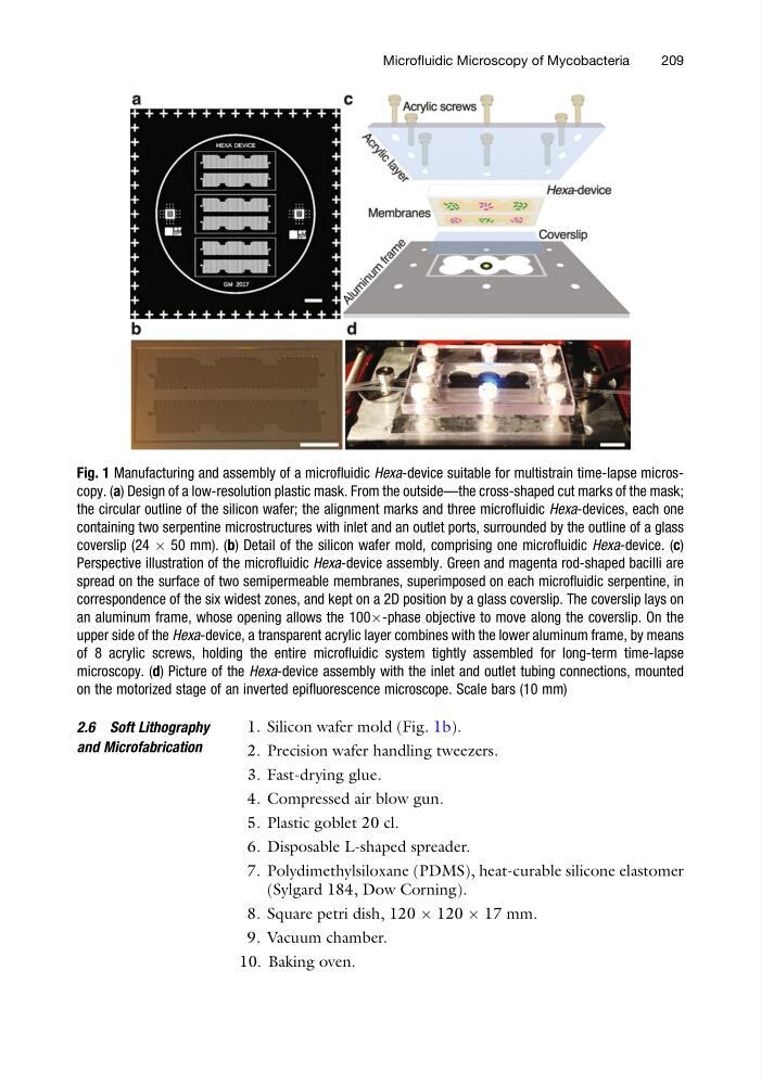
Fig. 1 Manufacturing and assembly of a microfluidic Hexa-device suitable for multistrain time-lapse microscopy. (a) Design of a low-resolution plastic mask. From the outside—the cross-shaped cut marks of the mask;
the circular outline of the silicon wafer; the alignment marks and three microfluidic Hexa-devices, each one
containing two serpentine microstructures with inlet and an outlet ports, surrounded by the outline of a glass
coverslip (24 Â 50 mm). (b) Detail of the silicon wafer mold, comprising one microfluidic Hexa-device. (c)
Perspective illustration of the microfluidic Hexa-device assembly. Green and magenta rod-shaped bacilli are
spread on the surface of two semipermeable membranes, superimposed on each microfluidic serpentine, in
correspondence of the six widest zones, and kept on a 2D position by a glass coverslip. The coverslip lays on
an aluminum frame, whose opening allows the 100Â-phase objective to move along the coverslip. On the
upper side of the Hexa-device, a transparent acrylic layer combines with the lower aluminum frame, by means
of 8 acrylic screws, holding the entire microfluidic system tightly assembled for long-term time-lapse
microscopy. (d) Picture of the Hexa-device assembly with the inlet and outlet tubing connections, mounted
on the motorized stage of an inverted epifluorescence microscope. Scale bars (10 mm)
Microfluidic Microscopy of Mycobacteria
209
and Microfabrication
1. Silicon wafer mold (Fig. 1b).
2. Precision wafer handling tweezers.
3. Fast-drying glue.
4. Compressed air blow gun.
5. Plastic goblet 20 cl.
6. Disposable L-shaped spreader.
7. Polydimethylsiloxane (PDMS), heat-curable silicone elastomer
(Sylgard 184, Dow Corning).
8. Square petri dish, 120 Â 120 Â 17 mm.
9. Vacuum chamber.
10. Baking oven.
Fig. 1 Manufacturing and assembly of a microfluidic Hexa-device suitable for multistrain time-lapse microscopy. (a) Design of a low-resolution plastic mask. From the outside—the cross-shaped cut marks of the mask;
the circular outline of the silicon wafer; the alignment marks and three microfluidic Hexa-devices, each one
containing two serpentine microstructures with inlet and an outlet ports, surrounded by the outline of a glass
coverslip (24 Â 50 mm). (b) Detail of the silicon wafer mold, comprising one microfluidic Hexa-device. (c)
Perspective illustration of the microfluidic Hexa-device assembly. Green and magenta rod-shaped bacilli are
spread on the surface of two semipermeable membranes, superimposed on each microfluidic serpentine, in
correspondence of the six widest zones, and kept on a 2D position by a glass coverslip. The coverslip lays on
an aluminum frame, whose opening allows the 100Â-phase objective to move along the coverslip. On the
upper side of the Hexa-device, a transparent acrylic layer combines with the lower aluminum frame, by means
of 8 acrylic screws, holding the entire microfluidic system tightly assembled for long-term time-lapse
microscopy. (d) Picture of the Hexa-device assembly with the inlet and outlet tubing connections, mounted
on the motorized stage of an inverted epifluorescence microscope. Scale bars (10 mm)
Microfluidic Microscopy of Mycobacteria
209