

11. Reverse back the 60% sucrose to the syringe at normal speed
and then disengage the flow cell by unscrewing the bottom of
the flow cell.
Place the sample, screw the bottom of the flow cell such that
the needle pierces through the tube and reaches about half an inch
into the gradient. Run the syringe pump (normal speed, 3 mL/
min, 20 s/tube) and collect the fractions while monitoring the OD
on the screen (see Note 14).
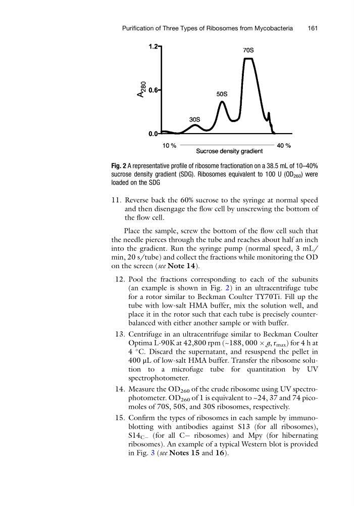
12. Pool the fractions corresponding to each of the subunits
(an example is shown in Fig. 2) in an ultracentrifuge tube
for a rotor similar to Beckman Coulter TY70Ti. Fill up the
tube with low-salt HMA buffer, mix the solution well, and
place it in the rotor such that each tube is precisely counterbalanced with either another sample or with buffer.
13. Centrifuge in an ultracentrifuge similar to Beckman Coulter
Optima L-90K at 42,800 rpm (~188, 000 Â g, r max ) for 4 h at
4
C. Discard the supernatant, and resuspend the pellet in
400 μL of low-salt HMA buffer. Transfer the ribosome solution to a microfuge tube for quantitation by UV
spectrophotometer.
14. Measure the OD 260 of the crude ribosome using UV spectrophotometer. OD 260 of 1 is equivalent to ~24, 37 and 74 picomoles of 70S, 50S, and 30S ribosomes, respectively.
15. Confirm the types of ribosomes in each sample by immunoblotting with antibodies against S13 (for all ribosomes),
S14 CÀ (for all CÀ ribosomes) and Mpy (for hibernating
ribosomes). An example of a typical Western blot is provided
in Fig. 3 (see Notes 15 and 16).
Fig. 2 A representative profile of ribosome fractionation on a 38.5 mL of 10–40%
sucrose density gradient (SDG). Ribosomes equivalent to 100 U (OD 260 ) were
loaded on the SDG
Purification of Three Types of Ribosomes from Mycobacteria
161
and then disengage the flow cell by unscrewing the bottom of
the flow cell.
Place the sample, screw the bottom of the flow cell such that
the needle pierces through the tube and reaches about half an inch
into the gradient. Run the syringe pump (normal speed, 3 mL/
min, 20 s/tube) and collect the fractions while monitoring the OD
on the screen (see Note 14).
12. Pool the fractions corresponding to each of the subunits
(an example is shown in Fig. 2) in an ultracentrifuge tube
for a rotor similar to Beckman Coulter TY70Ti. Fill up the
tube with low-salt HMA buffer, mix the solution well, and
place it in the rotor such that each tube is precisely counterbalanced with either another sample or with buffer.
13. Centrifuge in an ultracentrifuge similar to Beckman Coulter
Optima L-90K at 42,800 rpm (~188, 000 Â g, r max ) for 4 h at
4
C. Discard the supernatant, and resuspend the pellet in
400 μL of low-salt HMA buffer. Transfer the ribosome solution to a microfuge tube for quantitation by UV
spectrophotometer.
14. Measure the OD 260 of the crude ribosome using UV spectrophotometer. OD 260 of 1 is equivalent to ~24, 37 and 74 picomoles of 70S, 50S, and 30S ribosomes, respectively.
15. Confirm the types of ribosomes in each sample by immunoblotting with antibodies against S13 (for all ribosomes),
S14 CÀ (for all CÀ ribosomes) and Mpy (for hibernating
ribosomes). An example of a typical Western blot is provided
in Fig. 3 (see Notes 15 and 16).
Fig. 2 A representative profile of ribosome fractionation on a 38.5 mL of 10–40%
sucrose density gradient (SDG). Ribosomes equivalent to 100 U (OD 260 ) were
loaded on the SDG
Purification of Three Types of Ribosomes from Mycobacteria
161