

Processes 2018, 6,42
1. Introduction
Transcript variation at the single nucleotide level is increasingly being found to have widespread
occurrences within the transcriptome with fundamental roles in numerous biological processes
including development and disease. Specifically, several independent studies have reported that
there are hundreds of thousands of RNA editing sites catalyzed by the enzyme ADAR (adenosine
deaminase acting on RNA) within human mRNAs [1–6]. Editing via ADAR is characterized by the
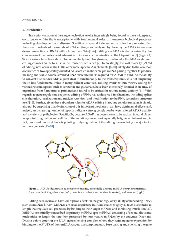
conversion of the nucleic acid adenosine to inosine via deamination at the C6 position [7] (Figure 1).
Since inosines have been shown to preferentially bind to cytosines, functionally the ADAR-catalyzed
editing changes an ‘A’ to a ‘G’ in the transcript sequence [7]. Interestingly, the vast majority (>99%)
of editing sites occur in the UTRs of primate-specific Alu elements [8–10], likely due to the common
occurrence of two oppositely oriented Alus located in the same pre-mRNA pairing together to produce
the long and stable double-stranded RNA structure that is required for ADAR to bind. As the ability
to convert nucleotides adds a great deal of functionality to the transcriptome, it is not surprising
that it has fundamental roles in many cellular activities. Editing events within mRNA coding for
various neuroreceptors, such as serotonin and glutamate, have been intensively detailed in an array of
organisms from flatworms to primates and found to be critical for routine neural activity [11]. With
regards to gene regulation, sequence editing of RNA has widespread implications, including splice
site alteration, localization and nuclear retention, and modification to the RNA secondary structure
itself [12]. Further, given these abundant roles for ADAR editing in routine cellular function, it should
also not be surprising that dysfunction of this important mechanism can have detrimental effects and,
indeed, an increasing number of reports indicate a strong correlation between altered ADAR activity
and a variety of pathologies. Specifically, because ADAR has been shown to be such an integral player
in apoptotic regulation and cellular differentiation, cancer is of especially heightened interest and, in
fact, more and more evidence is pointing to dysregulation of the editing process being a major factor
in tumorigenesis [13–18].
Figure 1. ADARs deaminate adenosine to inosine, potentially altering miRNA complementarities.
A cartoon depicting adenosine (left), deaminated adenosine (inosine, in center), and guanine (right).
Editing events can also have widespread effects on the gene regulatory ability of noncoding RNAs,
such as miRNAs [17,19]. MiRNAs are small regulatory RNA molecules roughly 20 to 23 nucleotides in
length that regulate cell processes by binding to their target mRNAs and inhibiting translation [20].
MiRNAs are initially transcribed as primary miRNAs (pri-miRNAs) consisting of several thousand
nucleotides in length that are then processed by into mature miRNAs by the enzymes Dicer and
Drosha before entering the RNAi gene silencing complex where they regulate gene expression by
binding to the 3 ′ UTR of their mRNA targets via complimentary base pairing and silencing the gene
131
1. Introduction
Transcript variation at the single nucleotide level is increasingly being found to have widespread
occurrences within the transcriptome with fundamental roles in numerous biological processes
including development and disease. Specifically, several independent studies have reported that
there are hundreds of thousands of RNA editing sites catalyzed by the enzyme ADAR (adenosine
deaminase acting on RNA) within human mRNAs [1–6]. Editing via ADAR is characterized by the
conversion of the nucleic acid adenosine to inosine via deamination at the C6 position [7] (Figure 1).
Since inosines have been shown to preferentially bind to cytosines, functionally the ADAR-catalyzed
editing changes an ‘A’ to a ‘G’ in the transcript sequence [7]. Interestingly, the vast majority (>99%)
of editing sites occur in the UTRs of primate-specific Alu elements [8–10], likely due to the common
occurrence of two oppositely oriented Alus located in the same pre-mRNA pairing together to produce
the long and stable double-stranded RNA structure that is required for ADAR to bind. As the ability
to convert nucleotides adds a great deal of functionality to the transcriptome, it is not surprising
that it has fundamental roles in many cellular activities. Editing events within mRNA coding for
various neuroreceptors, such as serotonin and glutamate, have been intensively detailed in an array of
organisms from flatworms to primates and found to be critical for routine neural activity [11]. With
regards to gene regulation, sequence editing of RNA has widespread implications, including splice
site alteration, localization and nuclear retention, and modification to the RNA secondary structure
itself [12]. Further, given these abundant roles for ADAR editing in routine cellular function, it should
also not be surprising that dysfunction of this important mechanism can have detrimental effects and,
indeed, an increasing number of reports indicate a strong correlation between altered ADAR activity
and a variety of pathologies. Specifically, because ADAR has been shown to be such an integral player
in apoptotic regulation and cellular differentiation, cancer is of especially heightened interest and, in
fact, more and more evidence is pointing to dysregulation of the editing process being a major factor
in tumorigenesis [13–18].
Figure 1. ADARs deaminate adenosine to inosine, potentially altering miRNA complementarities.
A cartoon depicting adenosine (left), deaminated adenosine (inosine, in center), and guanine (right).
Editing events can also have widespread effects on the gene regulatory ability of noncoding RNAs,
such as miRNAs [17,19]. MiRNAs are small regulatory RNA molecules roughly 20 to 23 nucleotides in
length that regulate cell processes by binding to their target mRNAs and inhibiting translation [20].
MiRNAs are initially transcribed as primary miRNAs (pri-miRNAs) consisting of several thousand
nucleotides in length that are then processed by into mature miRNAs by the enzymes Dicer and
Drosha before entering the RNAi gene silencing complex where they regulate gene expression by
binding to the 3 ′ UTR of their mRNA targets via complimentary base pairing and silencing the gene
131