

247
Craniofacial Development and Disorders
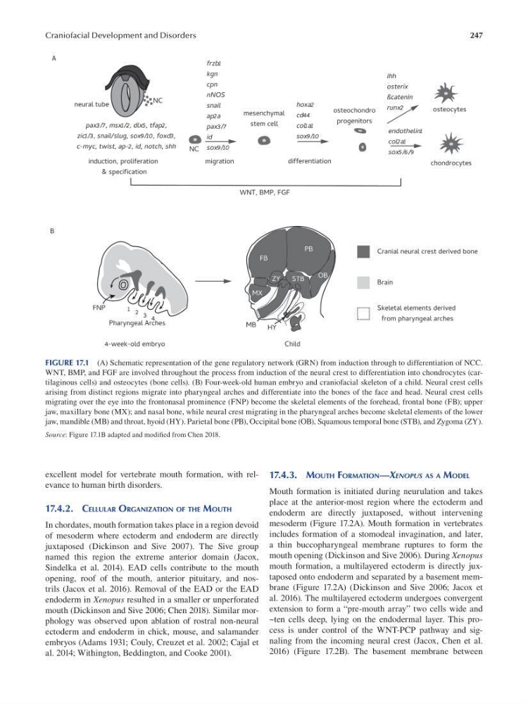
FIGURE 17.1 (A) Schematic representation of the gene regulatory network (GRN) from induction through to differentiation of NCC.
WNT, BMP, and FGF are involved throughout the process from induction of the neural crest to differentiation into chondrocytes (cartilaginous cells) and osteocytes (bone cells). (B) Four-week-old human embryo and craniofacial skeleton of a child. Neural crest cells
arising from distinct regions migrate into pharyngeal arches and differentiate into the bones of the face and head. Neural crest cells
migrating over the eye into the frontonasal prominence (FNP) become the skeletal elements of the forehead, frontal bone (FB); upper
jaw, maxillary bone (MX); and nasal bone, while neural crest migrating in the pharyngeal arches become skeletal elements of the lower
jaw, mandible (MB) and throat, hyoid (HY). Parietal bone (PB), Occipital bone (OB), Squamous temporal bone (STB), and Zygoma (ZY).
Source: Figure 17.1B adapted and modifed from Chen 2018.
excellent model for vertebrate mouth formation, with relevance to human birth disorders.
17.4.2. CELLULAR ORGANIZATION OF THE MOUTH
In chordates, mouth formation takes place in a region devoid
of mesoderm where ectoderm and endoderm are directly
juxtaposed (Dickinson and Sive 2007). The Sive group
named this region the extreme anterior domain (Jacox,
Sindelka et al. 2014). EAD cells contribute to the mouth
opening, roof of the mouth, anterior pituitary, and nostrils (Jacox et al. 2016). Removal of the EAD or the EAD
endoderm in Xenopus resulted in a smaller or unperforated
mouth (Dickinson and Sive 2006; Chen 2018). Similar morphology was observed upon ablation of rostral non-neural
ectoderm and endoderm in chick, mouse, and salamander
embryos (Adams 1931; Couly, Creuzet et al. 2002; Cajal et
al. 2014; Withington, Beddington, and Cooke 2001).
17.4.3. MOUTH FORMATION—XENOPUS AS A MODEL
Mouth formation is initiated during neurulation and takes
place at the anterior-most region where the ectoderm and
endoderm are directly juxtaposed, without intervening
mesoderm (Figure 17.2A). Mouth formation in vertebrates
includes formation of a stomodeal invagination, and later,
a thin buccopharyngeal membrane ruptures to form the
mouth opening (Dickinson and Sive 2006). During Xenopus
mouth formation, a multilayered ectoderm is directly juxtaposed onto endoderm and separated by a basement membrane (Figure 17.2A) (Dickinson and Sive 2006; Jacox et
al. 2016). The multilayered ectoderm undergoes convergent
extension to form a “pre-mouth array” two cells wide and
~ten cells deep, lying on the endodermal layer. This process is under control of the WNT-PCP pathway and signaling from the incoming neural crest (Jacox, Chen et al.
2016) (Figure 17.2B). The basement membrane between
Craniofacial Development and Disorders
FIGURE 17.1 (A) Schematic representation of the gene regulatory network (GRN) from induction through to differentiation of NCC.
WNT, BMP, and FGF are involved throughout the process from induction of the neural crest to differentiation into chondrocytes (cartilaginous cells) and osteocytes (bone cells). (B) Four-week-old human embryo and craniofacial skeleton of a child. Neural crest cells
arising from distinct regions migrate into pharyngeal arches and differentiate into the bones of the face and head. Neural crest cells
migrating over the eye into the frontonasal prominence (FNP) become the skeletal elements of the forehead, frontal bone (FB); upper
jaw, maxillary bone (MX); and nasal bone, while neural crest migrating in the pharyngeal arches become skeletal elements of the lower
jaw, mandible (MB) and throat, hyoid (HY). Parietal bone (PB), Occipital bone (OB), Squamous temporal bone (STB), and Zygoma (ZY).
Source: Figure 17.1B adapted and modifed from Chen 2018.
excellent model for vertebrate mouth formation, with relevance to human birth disorders.
17.4.2. CELLULAR ORGANIZATION OF THE MOUTH
In chordates, mouth formation takes place in a region devoid
of mesoderm where ectoderm and endoderm are directly
juxtaposed (Dickinson and Sive 2007). The Sive group
named this region the extreme anterior domain (Jacox,
Sindelka et al. 2014). EAD cells contribute to the mouth
opening, roof of the mouth, anterior pituitary, and nostrils (Jacox et al. 2016). Removal of the EAD or the EAD
endoderm in Xenopus resulted in a smaller or unperforated
mouth (Dickinson and Sive 2006; Chen 2018). Similar morphology was observed upon ablation of rostral non-neural
ectoderm and endoderm in chick, mouse, and salamander
embryos (Adams 1931; Couly, Creuzet et al. 2002; Cajal et
al. 2014; Withington, Beddington, and Cooke 2001).
17.4.3. MOUTH FORMATION—XENOPUS AS A MODEL
Mouth formation is initiated during neurulation and takes
place at the anterior-most region where the ectoderm and
endoderm are directly juxtaposed, without intervening
mesoderm (Figure 17.2A). Mouth formation in vertebrates
includes formation of a stomodeal invagination, and later,
a thin buccopharyngeal membrane ruptures to form the
mouth opening (Dickinson and Sive 2006). During Xenopus
mouth formation, a multilayered ectoderm is directly juxtaposed onto endoderm and separated by a basement membrane (Figure 17.2A) (Dickinson and Sive 2006; Jacox et
al. 2016). The multilayered ectoderm undergoes convergent
extension to form a “pre-mouth array” two cells wide and
~ten cells deep, lying on the endodermal layer. This process is under control of the WNT-PCP pathway and signaling from the incoming neural crest (Jacox, Chen et al.
2016) (Figure 17.2B). The basement membrane between