

230
Xenopus
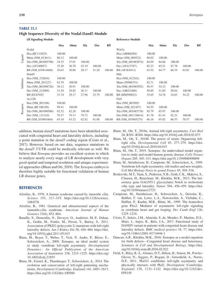
TABLE 15.1
High Sequence Diversity of the Nodal-Dand5 Module
LR Signaling Module
Reference Module
Hsa
Mmu
Xla
Dre
Bfl
Hsa
Mmu
Xla
Dre
Bfl
Nodal
Wnt3a
Hsa (BC112025)
100.00
Hsa (AB060284)
100.00
Mmu (NM_013611)
80.12
100.00
Mmu (NM_009522)
96.02
100.00
Xla (NM_001085796) 34.35
37.01
100.00
Xla (NM_001085874) 84.09
84.66
100.00
Dre (AY240027)
35.20
36.70
43.19
100.00
Dre (AY613787)
85.23
85.51
87.78
100.00
Bf (XM_035814606) 30.63
30.88
28.17
31.20
100.00 Bf (AF361013)
63.92
64.77
66.19
63.01
100.00
Dand5
Dkk
Hsa (NM_152654)
100.00
Hsa (NM_012242)
100.00
Mmu (NM_201227)
63.39
100.00
Mmu (JN966751)
82.71
100.00
Xla (NM_001098726) 30.12
30.91
100.00
Xla (NM_001085592) 56.57
54.12
100.00
Dre (NM_212969)
31.39
29.85
26.11
100.00
Dre (AB023488)
50.85
51.69
50.64
100.00
Bf (EU67025
31.74
29.17
27.96
23.79
100.00 Bf (HM590023)
35.65
34.76
34.63
34.22
100.00
Acvr2b
Frd1
Hsa (NM_001106)
100.00
Hsa (NM_003505)
100.00
Mmu (BC106189)
99.41
100.00
Mmu (NM_021457)
94.55
100.00
Xla (NM_001090580) 82.32
82.28
100.00
Xla (NM_001085738) 85.79
85.97
100.00
Dre (NM_131210)
79.37
79.13
79.72
100.00
Dre (NM_001130614) 81.78
81.41
82.21
100.00
Bf (XM_035801604) 63.10
63.22
62.82
61.88
100.00 Bf (XM_035805275) 66.18
65.82
68.53
70.57
100.00
Receptor
Inhibitor
Ligand
addition, human dand5 mutations have been identif ed associated with congenital heart and laterality defects, including
a point mutation in the dand5 coding region (Cristo et al.,
2017). However, based on our data, sequence mutations in
the dand5 3’UTR could be medically relevant as well. We
believe that Xenopus provides an excellent system in which
to analyze nearly every stage of LR development with very
good spatial and temporal resolution and unique experimental approaches (Blum and Ott, 2019). The Xenopus embryo is
therefore highly suitable for functional validation of human
LR disease genes.
REFERENCES
Afzelius, B., 1976. A human syndrome caused by immotile cilia.
Science 193, 317–319. https://doi.org/10.1126/science.
1084576
Afzelius, B., 1981. Genetical and ultrastructural aspects of the
immotile-cilia syndrome. American Journal of Human
Genetics 33(6), 852–864.
Bataille, S., Demoulin, N., Devuyst, O., Audrézet, M.-P., Dahan,
K., Godin, M., Fontès, M., Pirson, Y., Burtey, S., 2011.
Association of PKD2 (polycystin 2) mutations with left-right
laterality defects. Am J Kidney Dis 58, 456–460. https://doi.
org/10.1053/j.ajkd.2011.05.015
Blum, M., Beyer, T., Weber, T., Vick, P., Andre, P., Bitzer, E.,
Schweickert, A., 2009. Xenopus, an ideal model system
to study vertebrate left-right asymmetry. Developmental
Dynamics : An Offcial Publication of the American
Association of Anatomists 238, 1215–1225. https://doi.org/
10.1002/dvdy.21855
Blum, M., Feistel, K., Thumberger, T., Schweickert, A., 2014. The
evolution and conservation of left-right patterning mechanisms. Development (Cambridge, England) 141, 1603–1613.
https://doi.org/10.1242/dev.100560
Blum, M., Ott, T., 2018a. Animal left-right asymmetry. Curr Biol
28, R301–R304. https://doi.org/10.1016/j.cub.2018.02.073
Blum, M., Ott, T., 2018b. The power of strain: Organizing leftright cilia. Developmental Cell 45, 277–279. https://doi.
org/10.1016/j.devcel.2018.04.015
Blum, M., Ott, T., 2019. Xenopus: An undervalued model organism to study and model human genetic disease. Cells Tissues
Organs 205, 303–313. https://doi.org/10.1159/000490898
Blum, M., Steinbeisser, H., Campione, M., Schweickert, A., 1999.
Vertebrate left-right asymmetry: old studies and new insights.
Cell Mol Biology Noisy-le-grand France 45, 505–516.
Boskovski, M.T., Yuan, S., Pedersen, N.B., Goth, C.K., Makova, S.,
Clausen, H., Brueckner, M., Khokha, M.K., 2013. The heterotaxy gene GALNT11 glycosylates Notch to orchestrate
cilia type and laterality. Nature 504, 456–459. https://doi.
org/10.1038/nature12723
Campione, M., Steinbeisser, H., Schweickert, A., Deissler, K.,
Bebber, F. van, Lowe, L.A., Nowotschin, S., Viebahn, C.,
Haffter, P., Kuehn, M.R., Blum, M., 1999. The homeobox
gene Pitx2: Mediator of asymmetric left-right signaling
in vertebrate heart and gut looping. Dev Camb Engl 126,
1225–1234.
Cristo, F., Inácio, J.M., Almeida, S. de, Mendes, P., Martins, D.S.,
Maio, J., Anjos, R., Belo, J.A., 2017. Functional study of
DAND5 variant in patients with congenital heart disease and
laterality defects. BMC medical genetics 18, 77. https://doi.
org/10.1186/s12881-017-0444-1
Duncan, A.R., Khokha, M.K., 2016. Xenopus as a model organism
for birth defects—Congenital heart disease and heterotaxy.
Seminars in Cell and Developmental Biology. https://doi.
org/10.1016/j.semcdb.2016.02.022
Field, S., Riley, K.-L., Grimes, D.T., Hilton, H., Simon, M., PowlesGlover, N., Siggers, P., Bogani, D., Greenf eld, A., Norris,
D.P., 2011. Pkd1l1 establishes left-right asymmetry and
physically interacts with Pkd2. Development (Cambridge,
England) 138, 1131–1142. https://doi.org/10.1242/dev.
058149
Xenopus
TABLE 15.1
High Sequence Diversity of the Nodal-Dand5 Module
LR Signaling Module
Reference Module
Hsa
Mmu
Xla
Dre
Bfl
Hsa
Mmu
Xla
Dre
Bfl
Nodal
Wnt3a
Hsa (BC112025)
100.00
Hsa (AB060284)
100.00
Mmu (NM_013611)
80.12
100.00
Mmu (NM_009522)
96.02
100.00
Xla (NM_001085796) 34.35
37.01
100.00
Xla (NM_001085874) 84.09
84.66
100.00
Dre (AY240027)
35.20
36.70
43.19
100.00
Dre (AY613787)
85.23
85.51
87.78
100.00
Bf (XM_035814606) 30.63
30.88
28.17
31.20
100.00 Bf (AF361013)
63.92
64.77
66.19
63.01
100.00
Dand5
Dkk
Hsa (NM_152654)
100.00
Hsa (NM_012242)
100.00
Mmu (NM_201227)
63.39
100.00
Mmu (JN966751)
82.71
100.00
Xla (NM_001098726) 30.12
30.91
100.00
Xla (NM_001085592) 56.57
54.12
100.00
Dre (NM_212969)
31.39
29.85
26.11
100.00
Dre (AB023488)
50.85
51.69
50.64
100.00
Bf (EU67025
31.74
29.17
27.96
23.79
100.00 Bf (HM590023)
35.65
34.76
34.63
34.22
100.00
Acvr2b
Frd1
Hsa (NM_001106)
100.00
Hsa (NM_003505)
100.00
Mmu (BC106189)
99.41
100.00
Mmu (NM_021457)
94.55
100.00
Xla (NM_001090580) 82.32
82.28
100.00
Xla (NM_001085738) 85.79
85.97
100.00
Dre (NM_131210)
79.37
79.13
79.72
100.00
Dre (NM_001130614) 81.78
81.41
82.21
100.00
Bf (XM_035801604) 63.10
63.22
62.82
61.88
100.00 Bf (XM_035805275) 66.18
65.82
68.53
70.57
100.00
Receptor
Inhibitor
Ligand
addition, human dand5 mutations have been identif ed associated with congenital heart and laterality defects, including
a point mutation in the dand5 coding region (Cristo et al.,
2017). However, based on our data, sequence mutations in
the dand5 3’UTR could be medically relevant as well. We
believe that Xenopus provides an excellent system in which
to analyze nearly every stage of LR development with very
good spatial and temporal resolution and unique experimental approaches (Blum and Ott, 2019). The Xenopus embryo is
therefore highly suitable for functional validation of human
LR disease genes.
REFERENCES
Afzelius, B., 1976. A human syndrome caused by immotile cilia.
Science 193, 317–319. https://doi.org/10.1126/science.
1084576
Afzelius, B., 1981. Genetical and ultrastructural aspects of the
immotile-cilia syndrome. American Journal of Human
Genetics 33(6), 852–864.
Bataille, S., Demoulin, N., Devuyst, O., Audrézet, M.-P., Dahan,
K., Godin, M., Fontès, M., Pirson, Y., Burtey, S., 2011.
Association of PKD2 (polycystin 2) mutations with left-right
laterality defects. Am J Kidney Dis 58, 456–460. https://doi.
org/10.1053/j.ajkd.2011.05.015
Blum, M., Beyer, T., Weber, T., Vick, P., Andre, P., Bitzer, E.,
Schweickert, A., 2009. Xenopus, an ideal model system
to study vertebrate left-right asymmetry. Developmental
Dynamics : An Offcial Publication of the American
Association of Anatomists 238, 1215–1225. https://doi.org/
10.1002/dvdy.21855
Blum, M., Feistel, K., Thumberger, T., Schweickert, A., 2014. The
evolution and conservation of left-right patterning mechanisms. Development (Cambridge, England) 141, 1603–1613.
https://doi.org/10.1242/dev.100560
Blum, M., Ott, T., 2018a. Animal left-right asymmetry. Curr Biol
28, R301–R304. https://doi.org/10.1016/j.cub.2018.02.073
Blum, M., Ott, T., 2018b. The power of strain: Organizing leftright cilia. Developmental Cell 45, 277–279. https://doi.
org/10.1016/j.devcel.2018.04.015
Blum, M., Ott, T., 2019. Xenopus: An undervalued model organism to study and model human genetic disease. Cells Tissues
Organs 205, 303–313. https://doi.org/10.1159/000490898
Blum, M., Steinbeisser, H., Campione, M., Schweickert, A., 1999.
Vertebrate left-right asymmetry: old studies and new insights.
Cell Mol Biology Noisy-le-grand France 45, 505–516.
Boskovski, M.T., Yuan, S., Pedersen, N.B., Goth, C.K., Makova, S.,
Clausen, H., Brueckner, M., Khokha, M.K., 2013. The heterotaxy gene GALNT11 glycosylates Notch to orchestrate
cilia type and laterality. Nature 504, 456–459. https://doi.
org/10.1038/nature12723
Campione, M., Steinbeisser, H., Schweickert, A., Deissler, K.,
Bebber, F. van, Lowe, L.A., Nowotschin, S., Viebahn, C.,
Haffter, P., Kuehn, M.R., Blum, M., 1999. The homeobox
gene Pitx2: Mediator of asymmetric left-right signaling
in vertebrate heart and gut looping. Dev Camb Engl 126,
1225–1234.
Cristo, F., Inácio, J.M., Almeida, S. de, Mendes, P., Martins, D.S.,
Maio, J., Anjos, R., Belo, J.A., 2017. Functional study of
DAND5 variant in patients with congenital heart disease and
laterality defects. BMC medical genetics 18, 77. https://doi.
org/10.1186/s12881-017-0444-1
Duncan, A.R., Khokha, M.K., 2016. Xenopus as a model organism
for birth defects—Congenital heart disease and heterotaxy.
Seminars in Cell and Developmental Biology. https://doi.
org/10.1016/j.semcdb.2016.02.022
Field, S., Riley, K.-L., Grimes, D.T., Hilton, H., Simon, M., PowlesGlover, N., Siggers, P., Bogani, D., Greenf eld, A., Norris,
D.P., 2011. Pkd1l1 establishes left-right asymmetry and
physically interacts with Pkd2. Development (Cambridge,
England) 138, 1131–1142. https://doi.org/10.1242/dev.
058149