

212
Xenopus
cases, the re-integration was within 3MB of the donor locus
(Yergeau et al., 2011b).
Despite these promising initial results, no insertional
mutations were produced in these screens. However, a functionally disruptive mutation was discovered accidently when
an attempt was made to breed a Xenopus tropicalis line,
carrying a nkx2.5-GFP insert, to homozygosity (Abu-Daya
et al., 2011). Approximately 25% of metamorphic froglets
completely lacked forelimbs, including the scapula and clavicle. In situ hybridization showed that mutant tadpoles did
not express tbx5 in the prospective forelimb region, although
cardiac expression was unaffected. The integration site was
cloned by ligation-mediated PCR in the frst intron of the
nephronectin gene, a secreted ligand of Alpha8Beta1, which
was necessary for metanephros formation in mouse but had
not been implicated in limb generation prior to this point.
Early experiments thus showed the potential of using the
frog model for genetics experiments; however, before this
potential could be transformed into a major screening program as in other models, the landscape was changed by the
availability and low cost of large-scale sequencing together
with gene editing methods.
14.3.5. REVERSE GENETICS USING DSBREAKS
Reverse genetics really became feasible in Xenopus with the
introduction of new gene editing techniques that allowed targeted induction of double strand (ds) breaks in the genome
(Lei et al., 2013; Tandon et al., 2017). The frst study used
Zinc Finger Nucleases to demonstrate the possibility of targeted mutations in the Xenopus genome (Young et al., 2011).
The frst published studies with TALENS were in 2012, and
there have now been many different TALEN mutants created in both Xenopus species (2 in X. laevis and 14 in X.
tropicalis) (Ishibashi et al., 2012; Lei et al., 2012). Initially,
most of these studies analyzed F0 mutants with mosaic
mutations, but phenotypes were successfully observed.
The frst germline TALEN mutants generated were F1
compound heterozygotes; these X. tropicalis pax6 mutants
displayed phenotypes similar to human aniridia patients,
showing how Xenopus can be used to model human disease
(Nakayama et al., 2015). The f rst X. laevis germline mutant
was made using oocyte host transfer to improve mutation
rates in both tyr.L and tyr.S genes (Ratzan et al., 2017). This
paper showed that one can generate mutants effectively
in the allotetraploid X. laevis. The frst homozygous null
TALEN mutants were generated in the X. tropicalis protein
arginine histone methyltransferase 1 gene (prmt1 ) ( Shibata
et al., 2019). Prmt1 knockout mice die shortly after implantation, preventing functional analysis. Since Xenopus embryos
develop externally, prmt1 −/− embryos survive, allowing for
functional analysis, but show delayed growth after f ve days
and eventually die nine days later. Multiple genes can also
be mutated using TALENs. Knockout of rnf43 and znf3
together (but not individually) led to limb deformities in F0
X. tropicalis (Szenker-Ravi et al., 2018). TALEN use peaked
in terms of Xenopus publications in 2016, but already it had
been overtaken by a different method of making ds breaks.
Shortly after TALENs, the CRISPR-Cas technology
became available. The ease with which F0 Xenopus crispants can be made, the high level of indels caused, and the
strong penetrance of resulting phenotypes led this method to
become widely used in Xenopus. The f rst Xenopus CRISPR
mutants were published in 2013, and in the last f ve years,
over 50 different mutants have been published (Blitz et al.,
2013; Nakayama et al., 2013). Due to its diploidy and growth
at higher temperatures (where Cas is more effective), most
CRISPR mutants have been F0 mosaic mutants generated in
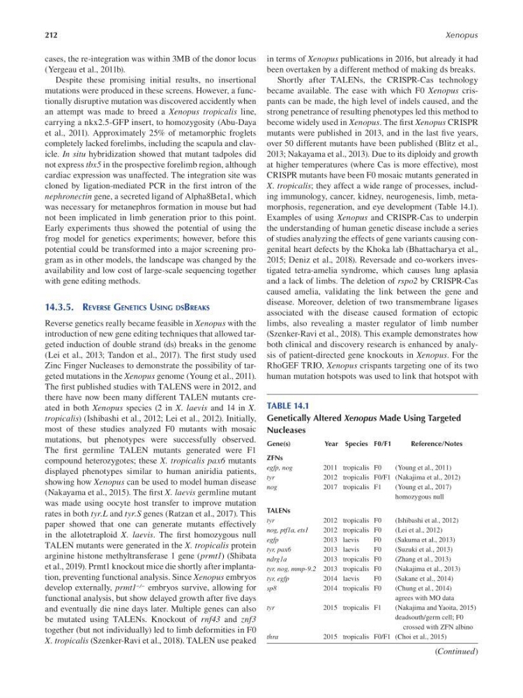
X. tropicalis; they affect a wide range of processes, including immunology, cancer, kidney, neurogenesis, limb, metamorphosis, regeneration, and eye development (Table 14.1).
Examples of using Xenopus and CRISPR-Cas to underpin
the understanding of human genetic disease include a series
of studies analyzing the effects of gene variants causing congenital heart defects by the Khoka lab (Bhattacharya et al.,
2015; Deniz et al., 2018). Reversade and co-workers investigated tetra-amelia syndrome, which causes lung aplasia
and a lack of limbs. The deletion of rspo2 by CRISPR-Cas
caused amelia, validating the link between the gene and
disease. Moreover, deletion of two transmembrane ligases
associated with the disease caused formation of ectopic
limbs, also revealing a master regulator of limb number
(Szenker-Ravi et al., 2018). This example demonstrates how
both clinical and discovery research is enhanced by analysis of patient-directed gene knockouts in Xenopus. For the
RhoGEF TRIO, Xenopus crispants targeting one of its two
human mutation hotspots was used to link that hotspot with
TABLE 14.1
Genetically Altered Xenopus Made Using Targeted
Nucleases
Gene(s)
Year Species F0/F1
Reference/Notes
ZFNs
egfp, nog
2011 tropicalis F0
( Young et al., 2011 )
tyr
2012 tropicalis F0/F1 ( Nakajima et al., 2012 )
nog
2017 tropicalis F1
( Young et al., 2017 )
homozygous null
TALENs
tyr
2012 tropicalis F0
( Ishibashi et al., 2012 )
nog, ptf1a, ets1
2012 tropicalis F0
( Lei et al., 2012 )
egfp
2013 laevis
F0
( Sakuma et al., 2013 )
tyr, pax6
2013 laevis
F0
( Suzuki et al., 2013 )
ndrg1a
2013 tropicalis F0
( Zhang et al., 2013 )
tyr, nog, mmp-9.2 2013 tropicalis F0
( Nakajima et al., 2013 )
tyr, egfp
2014 laevis
F0
( Sakane et al., 2014 )
sp8
2014 tropicalis F0
( Chung et al., 2014 )
agrees with MO data
tyr
2015 tropicalis F1
( Nakajima and Yaoita, 2015 )
deadsouth/germ cell; F0
crossed with ZFN albino
thra
2015 tropicalis F0/F1 ( Choi et al., 2015 )
(Continued)
Xenopus
cases, the re-integration was within 3MB of the donor locus
(Yergeau et al., 2011b).
Despite these promising initial results, no insertional
mutations were produced in these screens. However, a functionally disruptive mutation was discovered accidently when
an attempt was made to breed a Xenopus tropicalis line,
carrying a nkx2.5-GFP insert, to homozygosity (Abu-Daya
et al., 2011). Approximately 25% of metamorphic froglets
completely lacked forelimbs, including the scapula and clavicle. In situ hybridization showed that mutant tadpoles did
not express tbx5 in the prospective forelimb region, although
cardiac expression was unaffected. The integration site was
cloned by ligation-mediated PCR in the frst intron of the
nephronectin gene, a secreted ligand of Alpha8Beta1, which
was necessary for metanephros formation in mouse but had
not been implicated in limb generation prior to this point.
Early experiments thus showed the potential of using the
frog model for genetics experiments; however, before this
potential could be transformed into a major screening program as in other models, the landscape was changed by the
availability and low cost of large-scale sequencing together
with gene editing methods.
14.3.5. REVERSE GENETICS USING DSBREAKS
Reverse genetics really became feasible in Xenopus with the
introduction of new gene editing techniques that allowed targeted induction of double strand (ds) breaks in the genome
(Lei et al., 2013; Tandon et al., 2017). The frst study used
Zinc Finger Nucleases to demonstrate the possibility of targeted mutations in the Xenopus genome (Young et al., 2011).
The frst published studies with TALENS were in 2012, and
there have now been many different TALEN mutants created in both Xenopus species (2 in X. laevis and 14 in X.
tropicalis) (Ishibashi et al., 2012; Lei et al., 2012). Initially,
most of these studies analyzed F0 mutants with mosaic
mutations, but phenotypes were successfully observed.
The frst germline TALEN mutants generated were F1
compound heterozygotes; these X. tropicalis pax6 mutants
displayed phenotypes similar to human aniridia patients,
showing how Xenopus can be used to model human disease
(Nakayama et al., 2015). The f rst X. laevis germline mutant
was made using oocyte host transfer to improve mutation
rates in both tyr.L and tyr.S genes (Ratzan et al., 2017). This
paper showed that one can generate mutants effectively
in the allotetraploid X. laevis. The frst homozygous null
TALEN mutants were generated in the X. tropicalis protein
arginine histone methyltransferase 1 gene (prmt1 ) ( Shibata
et al., 2019). Prmt1 knockout mice die shortly after implantation, preventing functional analysis. Since Xenopus embryos
develop externally, prmt1 −/− embryos survive, allowing for
functional analysis, but show delayed growth after f ve days
and eventually die nine days later. Multiple genes can also
be mutated using TALENs. Knockout of rnf43 and znf3
together (but not individually) led to limb deformities in F0
X. tropicalis (Szenker-Ravi et al., 2018). TALEN use peaked
in terms of Xenopus publications in 2016, but already it had
been overtaken by a different method of making ds breaks.
Shortly after TALENs, the CRISPR-Cas technology
became available. The ease with which F0 Xenopus crispants can be made, the high level of indels caused, and the
strong penetrance of resulting phenotypes led this method to
become widely used in Xenopus. The f rst Xenopus CRISPR
mutants were published in 2013, and in the last f ve years,
over 50 different mutants have been published (Blitz et al.,
2013; Nakayama et al., 2013). Due to its diploidy and growth
at higher temperatures (where Cas is more effective), most
CRISPR mutants have been F0 mosaic mutants generated in
X. tropicalis; they affect a wide range of processes, including immunology, cancer, kidney, neurogenesis, limb, metamorphosis, regeneration, and eye development (Table 14.1).
Examples of using Xenopus and CRISPR-Cas to underpin
the understanding of human genetic disease include a series
of studies analyzing the effects of gene variants causing congenital heart defects by the Khoka lab (Bhattacharya et al.,
2015; Deniz et al., 2018). Reversade and co-workers investigated tetra-amelia syndrome, which causes lung aplasia
and a lack of limbs. The deletion of rspo2 by CRISPR-Cas
caused amelia, validating the link between the gene and
disease. Moreover, deletion of two transmembrane ligases
associated with the disease caused formation of ectopic
limbs, also revealing a master regulator of limb number
(Szenker-Ravi et al., 2018). This example demonstrates how
both clinical and discovery research is enhanced by analysis of patient-directed gene knockouts in Xenopus. For the
RhoGEF TRIO, Xenopus crispants targeting one of its two
human mutation hotspots was used to link that hotspot with
TABLE 14.1
Genetically Altered Xenopus Made Using Targeted
Nucleases
Gene(s)
Year Species F0/F1
Reference/Notes
ZFNs
egfp, nog
2011 tropicalis F0
( Young et al., 2011 )
tyr
2012 tropicalis F0/F1 ( Nakajima et al., 2012 )
nog
2017 tropicalis F1
( Young et al., 2017 )
homozygous null
TALENs
tyr
2012 tropicalis F0
( Ishibashi et al., 2012 )
nog, ptf1a, ets1
2012 tropicalis F0
( Lei et al., 2012 )
egfp
2013 laevis
F0
( Sakuma et al., 2013 )
tyr, pax6
2013 laevis
F0
( Suzuki et al., 2013 )
ndrg1a
2013 tropicalis F0
( Zhang et al., 2013 )
tyr, nog, mmp-9.2 2013 tropicalis F0
( Nakajima et al., 2013 )
tyr, egfp
2014 laevis
F0
( Sakane et al., 2014 )
sp8
2014 tropicalis F0
( Chung et al., 2014 )
agrees with MO data
tyr
2015 tropicalis F1
( Nakajima and Yaoita, 2015 )
deadsouth/germ cell; F0
crossed with ZFN albino
thra
2015 tropicalis F0/F1 ( Choi et al., 2015 )
(Continued)