

studies aimed at identifying and understanding the
origins of ‘source-specific’ D
14 C ; lipid ‘biomarker’
D
14 C ; compounds. Frequently, lipids from several
organic compound classes are studied within the
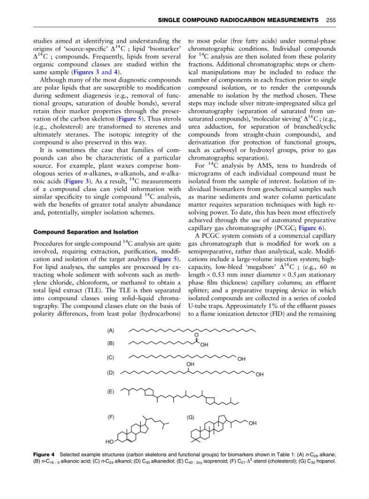
same sample (Figures 3 and 4).
Although many of the most diagnostic compounds
are polar lipids that are susceptible to modification
during sediment diagenesis (e.g., removal of functional groups, saturation of double bonds), several
retain their marker properties through the preservation of the carbon skeleton (Figure 5). Thus sterols
(e.g., cholesterol) are transformed to sterenes and
ultimately steranes. The isotopic integrity of the
compound is also preserved in this way.
It is sometimes the case that families of compounds can also be characteristic of a particular
source. For example, plant waxes comprise homologous series of n-alkanes, n-alkanols, and n-alkanoic acids (Figure 3). As a result,
14 C measurements
of a compound class can yield information with
similar specificity to single compound
14
C analysis,
with the benefits of greater total analyte abundance
and, potentially, simpler isolation schemes.
Compound Separation and Isolation
Procedures for single-compound
14
C analysis are quite
involved, requiring extraction, purification, modification and isolation of the target analytes (Figure 5).
For lipid analyses, the samples are processed by extracting whole sediment with solvents such as methylene chloride, chloroform, or methanol to obtain a
total lipid extract (TLE). The TLE is then separated
into compound classes using solid–liquid chromatography. The compound classes elute on the basis of
polarity differences, from least polar (hydrocarbons)
to most polar (free fatty acids) under normal-phase
chromatographic conditions. Individual compounds
for
14
C analysis are then isolated from these polarity
fractions. Additional chromatographic steps or chemical manipulations may be included to reduce the
number of components in each fraction prior to single
compound isolation, or to render the compounds
amenable to isolation by the method chosen. These
steps may include silver nitrate-impregnated silica gel
chromatography (separation of saturated from unsaturated compounds), ‘molecular sieving’ D
14 C ; (e.g.,
urea adduction, for separation of branched/cyclic
compounds from straight-chain compounds), and
derivatization (for protection of functional groups,
such as carboxyl or hydroxyl groups, prior to gas
chromatographic separation).
For
14 C analysis by AMS, tens to hundreds of
micrograms of each individual compound must be
isolated from the sample of interest. Isolation of individual biomarkers from geochemical samples such
as marine sediments and water column particulate
matter requires separation techniques with high resolving power. To date, this has been most effectively
achieved through the use of automated preparative
capillary gas chromatography (PCGC; Figure 6).
A PCGC system consists of a commercial capillary
gas chromatograph that is modified for work on a
semipreparative, rather than analytical, scale. Modifications include a large-volume injection system; highcapacity, low-bleed ‘megabore’ D
14 C ; (e.g., 60 m
length  0.53 mm inner diameter  0.5 mm stationary
phase film thickness) capillary columns; an effluent
splitter; and a preparative trapping device in which
isolated compounds are collected in a series of cooled
U-tube traps. Approximately 1% of the effluent passes
to a flame ionization detector (FID) and the remaining
O
OH
OH
OH
OH
HO
(E)
(G)
OH
(A)
(B)
(C)
(D)
(F)
Figure 4 Selected example structures (carbon skeletons and functional groups) for biomarkers shown in Table 1: (A) n-C 29 alkane;
(B) n-C 16 : 0 alkanoic acid; (C) n-C 24 alkanol; (D) C 30 alkanediol; (E) C 40 : 2cy isoprenoid; (F) C 27 D
5 -sterol (cholesterol); (G) C 32 hopanol.
SINGLE COMPOUND RADIOCARBON MEASUREMENTS 255
origins of ‘source-specific’ D
14 C ; lipid ‘biomarker’
D
14 C ; compounds. Frequently, lipids from several
organic compound classes are studied within the
same sample (Figures 3 and 4).
Although many of the most diagnostic compounds
are polar lipids that are susceptible to modification
during sediment diagenesis (e.g., removal of functional groups, saturation of double bonds), several
retain their marker properties through the preservation of the carbon skeleton (Figure 5). Thus sterols
(e.g., cholesterol) are transformed to sterenes and
ultimately steranes. The isotopic integrity of the
compound is also preserved in this way.
It is sometimes the case that families of compounds can also be characteristic of a particular
source. For example, plant waxes comprise homologous series of n-alkanes, n-alkanols, and n-alkanoic acids (Figure 3). As a result,
14 C measurements
of a compound class can yield information with
similar specificity to single compound
14
C analysis,
with the benefits of greater total analyte abundance
and, potentially, simpler isolation schemes.
Compound Separation and Isolation
Procedures for single-compound
14
C analysis are quite
involved, requiring extraction, purification, modification and isolation of the target analytes (Figure 5).
For lipid analyses, the samples are processed by extracting whole sediment with solvents such as methylene chloride, chloroform, or methanol to obtain a
total lipid extract (TLE). The TLE is then separated
into compound classes using solid–liquid chromatography. The compound classes elute on the basis of
polarity differences, from least polar (hydrocarbons)
to most polar (free fatty acids) under normal-phase
chromatographic conditions. Individual compounds
for
14
C analysis are then isolated from these polarity
fractions. Additional chromatographic steps or chemical manipulations may be included to reduce the
number of components in each fraction prior to single
compound isolation, or to render the compounds
amenable to isolation by the method chosen. These
steps may include silver nitrate-impregnated silica gel
chromatography (separation of saturated from unsaturated compounds), ‘molecular sieving’ D
14 C ; (e.g.,
urea adduction, for separation of branched/cyclic
compounds from straight-chain compounds), and
derivatization (for protection of functional groups,
such as carboxyl or hydroxyl groups, prior to gas
chromatographic separation).
For
14 C analysis by AMS, tens to hundreds of
micrograms of each individual compound must be
isolated from the sample of interest. Isolation of individual biomarkers from geochemical samples such
as marine sediments and water column particulate
matter requires separation techniques with high resolving power. To date, this has been most effectively
achieved through the use of automated preparative
capillary gas chromatography (PCGC; Figure 6).
A PCGC system consists of a commercial capillary
gas chromatograph that is modified for work on a
semipreparative, rather than analytical, scale. Modifications include a large-volume injection system; highcapacity, low-bleed ‘megabore’ D
14 C ; (e.g., 60 m
length  0.53 mm inner diameter  0.5 mm stationary
phase film thickness) capillary columns; an effluent
splitter; and a preparative trapping device in which
isolated compounds are collected in a series of cooled
U-tube traps. Approximately 1% of the effluent passes
to a flame ionization detector (FID) and the remaining
O
OH
OH
OH
OH
HO
(E)
(G)
OH
(A)
(B)
(C)
(D)
(F)
Figure 4 Selected example structures (carbon skeletons and functional groups) for biomarkers shown in Table 1: (A) n-C 29 alkane;
(B) n-C 16 : 0 alkanoic acid; (C) n-C 24 alkanol; (D) C 30 alkanediol; (E) C 40 : 2cy isoprenoid; (F) C 27 D
5 -sterol (cholesterol); (G) C 32 hopanol.
SINGLE COMPOUND RADIOCARBON MEASUREMENTS 255