

9
TIME (h)
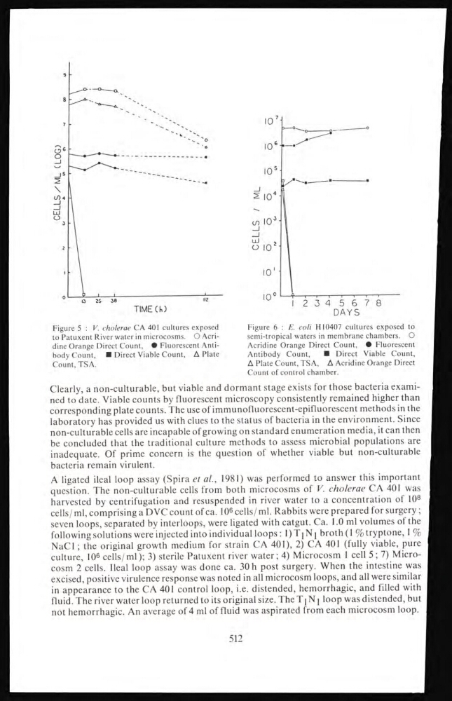
Figure 5 : V. cholerae CA 401 cultures exposed
to Patuxent River water in microcosms. O Acridine Orange Direct Count, • Fluorescent Antibody Count, ■ Direct Viable Count, A Plate
Count, TSA.
Figure 6 : E. coli H10407 cultures exposed to
semi-tropical waters in membrane chambers. O
Acridine Orange Direct Count, • Fluorescent
Antibody Count,
■ Direct Viable Count,
A Plate Count, TSA, A Acridine Orange Direct
Count of control chamber.
Clearly, a non-culturable, but viable and dormant stage exists for those bacteria examined to date. Viable counts by fluorescent microscopy consistently remained higher than
corresponding plate counts. The use of immunofluorescent-epifluorescent methods in the
laboratory has provided us with clues to the status of bacteria in the environment. Since
non-culturable cells are incapable of growing on standard enumeration media, it can then
be concluded that the traditional culture methods to assess microbial populations are
inadequate. Of prime concern is the question of whether viable but non-culturable
bacteria remain virulent.
A ligated ileal loop assay (Spira et al., 1981) was performed to answer this important
question. The non-culturable cells from both microcosms of V. cholerae CA 401 was
harvested by centrifugation and resuspended in river water to a concentration of 108
cells/ ml, comprising a DVC count of ca. 10 6 cells/ ml. Rabbits were prepared for surgery ;
seven loops, separated by interloops, were ligated with catgut. Ca. 1.0 ml volumes of the
following solutions were injected into individual loops: 1 ) T 1 N 1 broth (1 %tryptone, 1 %
NaCl ; the original growth medium for strain CA 401), 2) CA 401 (fully viable, pure
culture, 10
6 cells/ml); 3) sterile Patuxent river water; 4) Microcosm 1 cell 5; 7) Microcosm 2 cells. Ileal loop assay was done ca. 30 h post surgery. When the intestine was
excised, positive virulence response was noted in all microcosm loops, and all were similar
in appearance to the CA 401 control loop, i.e. distended, hemorrhagic, and filled with
fluid. The river water loop returned to its original size. The T 1
N 1
loop was distended, but
not hemorrhagic. An average of 4 ml of fluid was aspirated from each microcosm loop.
512
TIME (h)
Figure 5 : V. cholerae CA 401 cultures exposed
to Patuxent River water in microcosms. O Acridine Orange Direct Count, • Fluorescent Antibody Count, ■ Direct Viable Count, A Plate
Count, TSA.
Figure 6 : E. coli H10407 cultures exposed to
semi-tropical waters in membrane chambers. O
Acridine Orange Direct Count, • Fluorescent
Antibody Count,
■ Direct Viable Count,
A Plate Count, TSA, A Acridine Orange Direct
Count of control chamber.
Clearly, a non-culturable, but viable and dormant stage exists for those bacteria examined to date. Viable counts by fluorescent microscopy consistently remained higher than
corresponding plate counts. The use of immunofluorescent-epifluorescent methods in the
laboratory has provided us with clues to the status of bacteria in the environment. Since
non-culturable cells are incapable of growing on standard enumeration media, it can then
be concluded that the traditional culture methods to assess microbial populations are
inadequate. Of prime concern is the question of whether viable but non-culturable
bacteria remain virulent.
A ligated ileal loop assay (Spira et al., 1981) was performed to answer this important
question. The non-culturable cells from both microcosms of V. cholerae CA 401 was
harvested by centrifugation and resuspended in river water to a concentration of 108
cells/ ml, comprising a DVC count of ca. 10 6 cells/ ml. Rabbits were prepared for surgery ;
seven loops, separated by interloops, were ligated with catgut. Ca. 1.0 ml volumes of the
following solutions were injected into individual loops: 1 ) T 1 N 1 broth (1 %tryptone, 1 %
NaCl ; the original growth medium for strain CA 401), 2) CA 401 (fully viable, pure
culture, 10
6 cells/ml); 3) sterile Patuxent river water; 4) Microcosm 1 cell 5; 7) Microcosm 2 cells. Ileal loop assay was done ca. 30 h post surgery. When the intestine was
excised, positive virulence response was noted in all microcosm loops, and all were similar
in appearance to the CA 401 control loop, i.e. distended, hemorrhagic, and filled with
fluid. The river water loop returned to its original size. The T 1
N 1
loop was distended, but
not hemorrhagic. An average of 4 ml of fluid was aspirated from each microcosm loop.
512