

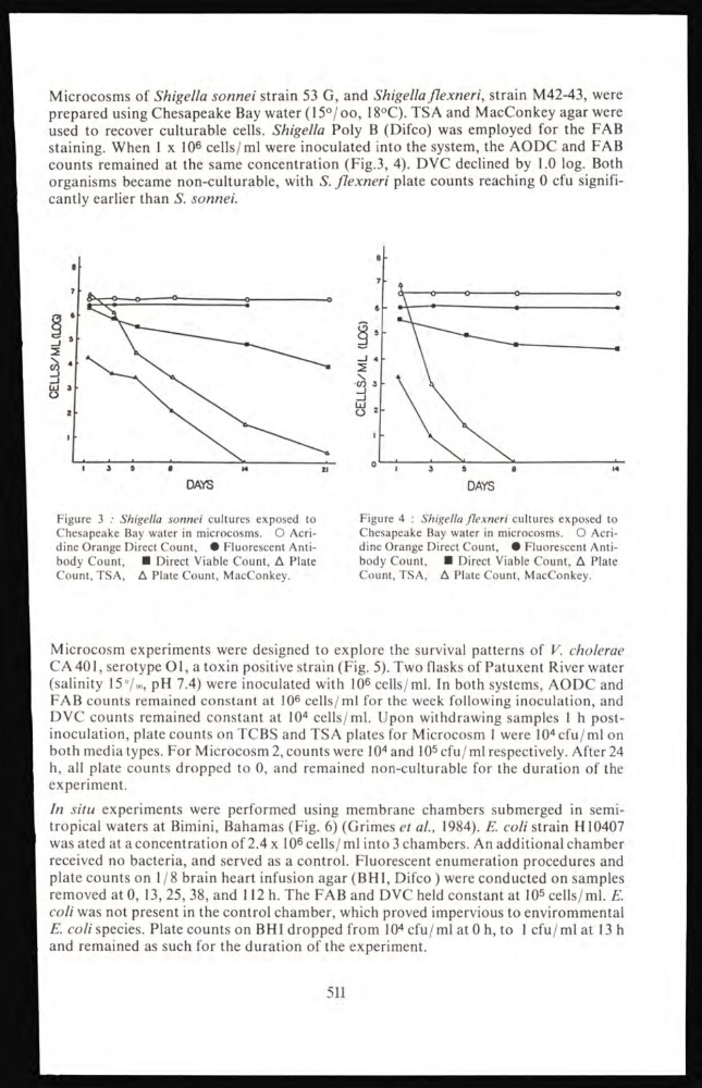
Microcosms of Shigella sonnei strain 53 G, and Shigella flexneri, strain M42-43, were
prepared using Chesapeake Bay water (15‰, 18°C). TSA and MacConkey agar were
used to recover culturable cells. Shigella Poly B (Difco) was employed for the FAB
staining. When 1 x 10 6 cells/ml were inoculated into the system, the AODC and FAB
counts remained at the same concentration (Fig.3, 4). DVC declined by 1.0 log. Both
organisms became non-culturable, with S. flexneri plate counts reaching 0 cfu significantly earlier than S. sonnei.
Figure 3 : Shigella sonnei cultures exposed to
Chesapeake Bay water in microcosms. O Acridine Orange Direct Count, • Fluorescent Antibody Count, ■ Direct Viable Count, A Plate
Count, TSA, A Plate Count, MacConkey.
Figure 4 : Shigella flexneri cultures exposed to
Chesapeake Bay water in microcosms. O Acridine Orange Direct Count, • Fluorescent Antibody Count,
■ Direct Viable Count, A Plate
Count, TSA, A Plate Count, MacConkey.
Microcosm experiments were designed to explore the survival patterns of V. cholerae
CA 401, serotype 01, a toxin positive strain (Fig. 5). Two flasks of Patuxent River water
(salinity 15‰, pH 7.4) were inoculated with 10
6 cells/ml. In both systems, AODC and
FAB counts remained constant at 10 6 cells/ml for the week following inoculation, and
DVC counts remained constant at 10
4 cells/ml. Upon withdrawing samples 1 h postinoculation, plate counts on TCBS and TSA plates for Microcosm 1 were 10
4 cfu/ml on
both media types. For Microcosm 2, counts were 10
4 and 10
5 cfu/ml respectively. After 24
h, all plate counts dropped to 0, and remained non-culturable for the duration of the
experiment.
In situ experiments were performed using membrane chambers submerged in semitropical waters at Bimini, Bahamas (Fig. 6) (Grimes et ai, 1984). E. coli strain H10407
was ated at a concentration of 2.4 x 10 6 cells/ml into 3 chambers. An additional chamber
received no bacteria, and served as a control. Fluorescent enumeration procedures and
plate counts on 1/8 brain heart infusion agar (BHI, Difco ) were conducted on samples
removed at 0, 13, 25, 38, and 112 h. The FAB and DVC held constant at 10 5 cells/ml. E.
coli was not present in the control chamber, which proved impervious to envirommental
E. coli species. Plate counts on BHI dropped from 10
4 cfu/ml at 0 h, to 1 cfu/ml at 13 h
and remained as such for the duration of the experiment.
511
prepared using Chesapeake Bay water (15‰, 18°C). TSA and MacConkey agar were
used to recover culturable cells. Shigella Poly B (Difco) was employed for the FAB
staining. When 1 x 10 6 cells/ml were inoculated into the system, the AODC and FAB
counts remained at the same concentration (Fig.3, 4). DVC declined by 1.0 log. Both
organisms became non-culturable, with S. flexneri plate counts reaching 0 cfu significantly earlier than S. sonnei.
Figure 3 : Shigella sonnei cultures exposed to
Chesapeake Bay water in microcosms. O Acridine Orange Direct Count, • Fluorescent Antibody Count, ■ Direct Viable Count, A Plate
Count, TSA, A Plate Count, MacConkey.
Figure 4 : Shigella flexneri cultures exposed to
Chesapeake Bay water in microcosms. O Acridine Orange Direct Count, • Fluorescent Antibody Count,
■ Direct Viable Count, A Plate
Count, TSA, A Plate Count, MacConkey.
Microcosm experiments were designed to explore the survival patterns of V. cholerae
CA 401, serotype 01, a toxin positive strain (Fig. 5). Two flasks of Patuxent River water
(salinity 15‰, pH 7.4) were inoculated with 10
6 cells/ml. In both systems, AODC and
FAB counts remained constant at 10 6 cells/ml for the week following inoculation, and
DVC counts remained constant at 10
4 cells/ml. Upon withdrawing samples 1 h postinoculation, plate counts on TCBS and TSA plates for Microcosm 1 were 10
4 cfu/ml on
both media types. For Microcosm 2, counts were 10
4 and 10
5 cfu/ml respectively. After 24
h, all plate counts dropped to 0, and remained non-culturable for the duration of the
experiment.
In situ experiments were performed using membrane chambers submerged in semitropical waters at Bimini, Bahamas (Fig. 6) (Grimes et ai, 1984). E. coli strain H10407
was ated at a concentration of 2.4 x 10 6 cells/ml into 3 chambers. An additional chamber
received no bacteria, and served as a control. Fluorescent enumeration procedures and
plate counts on 1/8 brain heart infusion agar (BHI, Difco ) were conducted on samples
removed at 0, 13, 25, 38, and 112 h. The FAB and DVC held constant at 10 5 cells/ml. E.
coli was not present in the control chamber, which proved impervious to envirommental
E. coli species. Plate counts on BHI dropped from 10
4 cfu/ml at 0 h, to 1 cfu/ml at 13 h
and remained as such for the duration of the experiment.
511