

MATERIAL AND METHODS
Specimens of Aplysia benedicti and Bullia tranquebarica were collected from the mouth
of the Vellar estuary and the sandy beach of the Bay of Bengal at Porto Novo, from June
to August, 1982 when these animals were plentiful.
Live animals were brought to the laboratory in polythene buckets, washed several times
with sterile sea water to prevent contamination from shell surface and mantle fluid,and
the gut of the animals were aseptically removed. The tissues adhering to the gut were
carefully removed using a sterile forceps and the gut homogenized in 100 ml of sterile sea
water. Serial dilutions were made from the homogenate and spread into petriplates
containing sea water complete (SWC) agar medium (Ruby and Nealson, 1978). The plates
were incubated in dark at room temperature (28°C) for 24 hrs following which the total
CFU (Colony Forming Units) and luminous CFU were counted. Single, bright luminous
colonies were picked randomly from the petriplates and maintained in SWC agar slants at
room temperature. The scheme of Nealson ( 1978) was followed for the identification of
the isolates.
The method of Ruby and Nealson (1978) was followed for screening luminous bacteria
from the water. The luminous bacteria from the sediment were isolated by the serial
dilution-spread-plate technique as in the case of molluscan gut.
The bacterial isolates were subjected to various regimes of temperature, sodium chloride
concentration and pH. Sodium chloride concentration and pH tolerance studies were
conducted at room temperature (28°C). Growth of the isolates was monitored by measuring the turbidity of the cultures at 550 nm in a Lumetron Colorimeter (Model N° 401,
photovolt Inc., New York). The assay of Reissig et al. (1955) was employed to determine
the chitinolytic activity of the isolates.
Animals
Collection period
Salinity
(%)
Oxygen
(ml/1)
Temp
(°C)
PH
A. benedicti
June, 1982
31.0
3.0
33
7.3
July
21.5
4.0
30
7.4
August
30.5
4.0
30
7.6
B. tranquebarica
June, 1982
31.5
3.2
33
7.4
July
30.5
3.9
29
7.3
August
31.8
3.8
28
7.4
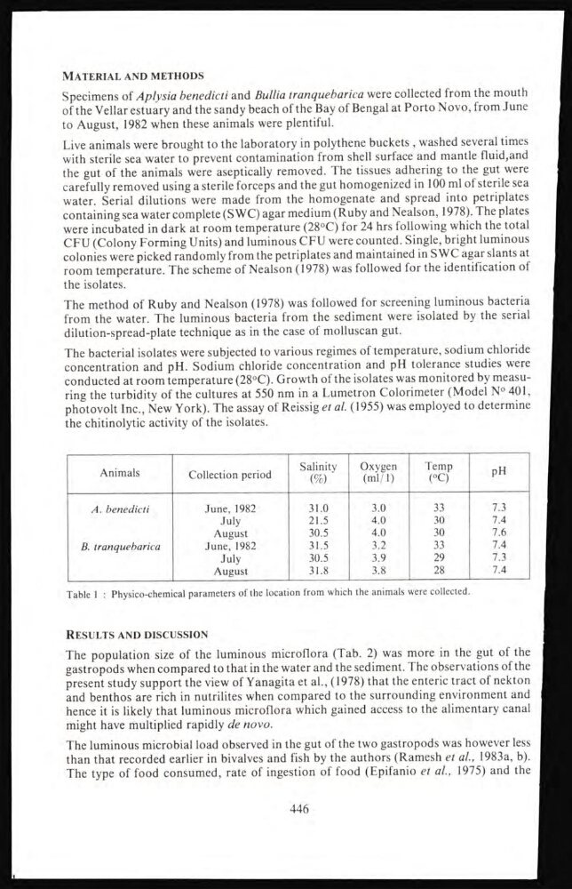
Table 1 : Physico-chemical parameters of the location from which the animals were collected.
RESULTS AND DISCUSSION
The population size of the luminous microflora (Tab. 2) was more in the gut of the
gastropods when compared to that in the water and the sediment. The observations of the
present study support the view of Yanagita et al., (1978) that the enteric tract of nekton
and benthos are rich in nutrilites when compared to the surrounding environment and
hence it is likely that luminous microflora which gained access to the alimentary canal
might have multiplied rapidly de novo.
The luminous microbial load observed in the gut of the two gastropods was however less
than that recorded earlier in bivalves and fish by the authors (Ramesh et al., 1983a, b).
The type of food consumed, rate of ingestion of food (Epifanio et al., 1975) and the
446
Specimens of Aplysia benedicti and Bullia tranquebarica were collected from the mouth
of the Vellar estuary and the sandy beach of the Bay of Bengal at Porto Novo, from June
to August, 1982 when these animals were plentiful.
Live animals were brought to the laboratory in polythene buckets, washed several times
with sterile sea water to prevent contamination from shell surface and mantle fluid,and
the gut of the animals were aseptically removed. The tissues adhering to the gut were
carefully removed using a sterile forceps and the gut homogenized in 100 ml of sterile sea
water. Serial dilutions were made from the homogenate and spread into petriplates
containing sea water complete (SWC) agar medium (Ruby and Nealson, 1978). The plates
were incubated in dark at room temperature (28°C) for 24 hrs following which the total
CFU (Colony Forming Units) and luminous CFU were counted. Single, bright luminous
colonies were picked randomly from the petriplates and maintained in SWC agar slants at
room temperature. The scheme of Nealson ( 1978) was followed for the identification of
the isolates.
The method of Ruby and Nealson (1978) was followed for screening luminous bacteria
from the water. The luminous bacteria from the sediment were isolated by the serial
dilution-spread-plate technique as in the case of molluscan gut.
The bacterial isolates were subjected to various regimes of temperature, sodium chloride
concentration and pH. Sodium chloride concentration and pH tolerance studies were
conducted at room temperature (28°C). Growth of the isolates was monitored by measuring the turbidity of the cultures at 550 nm in a Lumetron Colorimeter (Model N° 401,
photovolt Inc., New York). The assay of Reissig et al. (1955) was employed to determine
the chitinolytic activity of the isolates.
Animals
Collection period
Salinity
(%)
Oxygen
(ml/1)
Temp
(°C)
PH
A. benedicti
June, 1982
31.0
3.0
33
7.3
July
21.5
4.0
30
7.4
August
30.5
4.0
30
7.6
B. tranquebarica
June, 1982
31.5
3.2
33
7.4
July
30.5
3.9
29
7.3
August
31.8
3.8
28
7.4
Table 1 : Physico-chemical parameters of the location from which the animals were collected.
RESULTS AND DISCUSSION
The population size of the luminous microflora (Tab. 2) was more in the gut of the
gastropods when compared to that in the water and the sediment. The observations of the
present study support the view of Yanagita et al., (1978) that the enteric tract of nekton
and benthos are rich in nutrilites when compared to the surrounding environment and
hence it is likely that luminous microflora which gained access to the alimentary canal
might have multiplied rapidly de novo.
The luminous microbial load observed in the gut of the two gastropods was however less
than that recorded earlier in bivalves and fish by the authors (Ramesh et al., 1983a, b).
The type of food consumed, rate of ingestion of food (Epifanio et al., 1975) and the
446