

RESULTS AND DISCUSSION
Some examples from our work on extracellular enzymatic activity of marine bacteria are
provided here to demonstrate the applicability of the new technique for ecological as well
as physiological field studies. It should be premised that photographic documentation of
the results is not fully satisfactory due to the effects of long-wave UV radiation and the
dark blue background emission of the filter paper disks treated with MUF-substrate.
Furthermore, light emission from the HBO microscope lamp used here was not evenly
distributed over the total area of the filter paper. Light scattering did not fully prevent this
effect because it is always associated with a certain degree of light extinction. All these
implications are, however, not inherent problems for the visual observations of enzymatic
reactions on the filter paper disks. For the human eye the background (filter paper)
appears nearly black and the reactive spots glow intensively light blue or are simply
bright. Colonies lacking extracellular enzymatic activity do not provoke any MUF
fluorescence on the filter paper. Moreover, the somewhat irregular excitation irradiance
from the UV-lamp is not visible to the unaided eye.
Extracellular enzymatic activities of standard ZoBell agar plates
Inoculated with water from the Kiel Fjord, a ZoBell agar plate, as it is used to obtain
standard colony counts, is presented in Fig. 2a. Fig. 2b shows the filter paper which was
soaked with MUF-leucine and exposed to the agar plate colonies for 3 mn. Spots of
enzymatic reaction become visible with long-wave UV irradiation. Even those spots
caused by very small colonies are clearly recognizable. Two relatively large colonies did
not exhibit protease activity in the sense of MUF-leucine breakdown.
Extracellular enzymatic activity from selected colonies
7 agar plates were inoculated with 10 randomly selected colonies from standard ZoBell
agar plates. One example is given in Fig. 3a. Some enzymatic activities (protease,
phosphatase, lipase) are documented with photographs (Fig. 3b-d). The complete results
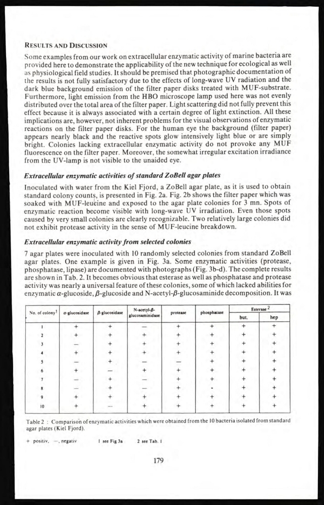
are shown in Tab. 2. It becomes obvious that esterase as well as phosphatase and protease
activity was nearly a universal feature of these colonies, some of which lacked abilities for
enzymatic α-glucoside, β-glucoside and N-acetyl-β-glucosaminide decomposition. It was
No. of colony 1
α-glucosidase
β-glucosidase
N-acetyl-βglucosaminidase
protease
phosphatase
Esterase 2
but.
hep
1
+
+
—
4+
+
42
+
+
+
+
+
+
+
3
—
+
+
+
+
+
+
4
+
+
+
+
+
4+
5
—
+
—
—
+
+
+
6
+
—
+
+
+
+
+
7
—
+
—
+
+
+
+
8
—
+
—
+
-
+
+
9
+
+
+
+
+
+
+
10
+
-
+
+
+
+
+
Table 2 : Comparison of enzymatic activities which were obtained from the 10 bacteria isolated from standard
agar plates (Kiel Fjord).
+ positiv. —. negativ
1 see Fig 3a
2 see Tab. I
179
Some examples from our work on extracellular enzymatic activity of marine bacteria are
provided here to demonstrate the applicability of the new technique for ecological as well
as physiological field studies. It should be premised that photographic documentation of
the results is not fully satisfactory due to the effects of long-wave UV radiation and the
dark blue background emission of the filter paper disks treated with MUF-substrate.
Furthermore, light emission from the HBO microscope lamp used here was not evenly
distributed over the total area of the filter paper. Light scattering did not fully prevent this
effect because it is always associated with a certain degree of light extinction. All these
implications are, however, not inherent problems for the visual observations of enzymatic
reactions on the filter paper disks. For the human eye the background (filter paper)
appears nearly black and the reactive spots glow intensively light blue or are simply
bright. Colonies lacking extracellular enzymatic activity do not provoke any MUF
fluorescence on the filter paper. Moreover, the somewhat irregular excitation irradiance
from the UV-lamp is not visible to the unaided eye.
Extracellular enzymatic activities of standard ZoBell agar plates
Inoculated with water from the Kiel Fjord, a ZoBell agar plate, as it is used to obtain
standard colony counts, is presented in Fig. 2a. Fig. 2b shows the filter paper which was
soaked with MUF-leucine and exposed to the agar plate colonies for 3 mn. Spots of
enzymatic reaction become visible with long-wave UV irradiation. Even those spots
caused by very small colonies are clearly recognizable. Two relatively large colonies did
not exhibit protease activity in the sense of MUF-leucine breakdown.
Extracellular enzymatic activity from selected colonies
7 agar plates were inoculated with 10 randomly selected colonies from standard ZoBell
agar plates. One example is given in Fig. 3a. Some enzymatic activities (protease,
phosphatase, lipase) are documented with photographs (Fig. 3b-d). The complete results
are shown in Tab. 2. It becomes obvious that esterase as well as phosphatase and protease
activity was nearly a universal feature of these colonies, some of which lacked abilities for
enzymatic α-glucoside, β-glucoside and N-acetyl-β-glucosaminide decomposition. It was
No. of colony 1
α-glucosidase
β-glucosidase
N-acetyl-βglucosaminidase
protease
phosphatase
Esterase 2
but.
hep
1
+
+
—
4+
+
42
+
+
+
+
+
+
+
3
—
+
+
+
+
+
+
4
+
+
+
+
+
4+
5
—
+
—
—
+
+
+
6
+
—
+
+
+
+
+
7
—
+
—
+
+
+
+
8
—
+
—
+
-
+
+
9
+
+
+
+
+
+
+
10
+
-
+
+
+
+
+
Table 2 : Comparison of enzymatic activities which were obtained from the 10 bacteria isolated from standard
agar plates (Kiel Fjord).
+ positiv. —. negativ
1 see Fig 3a
2 see Tab. I
179