

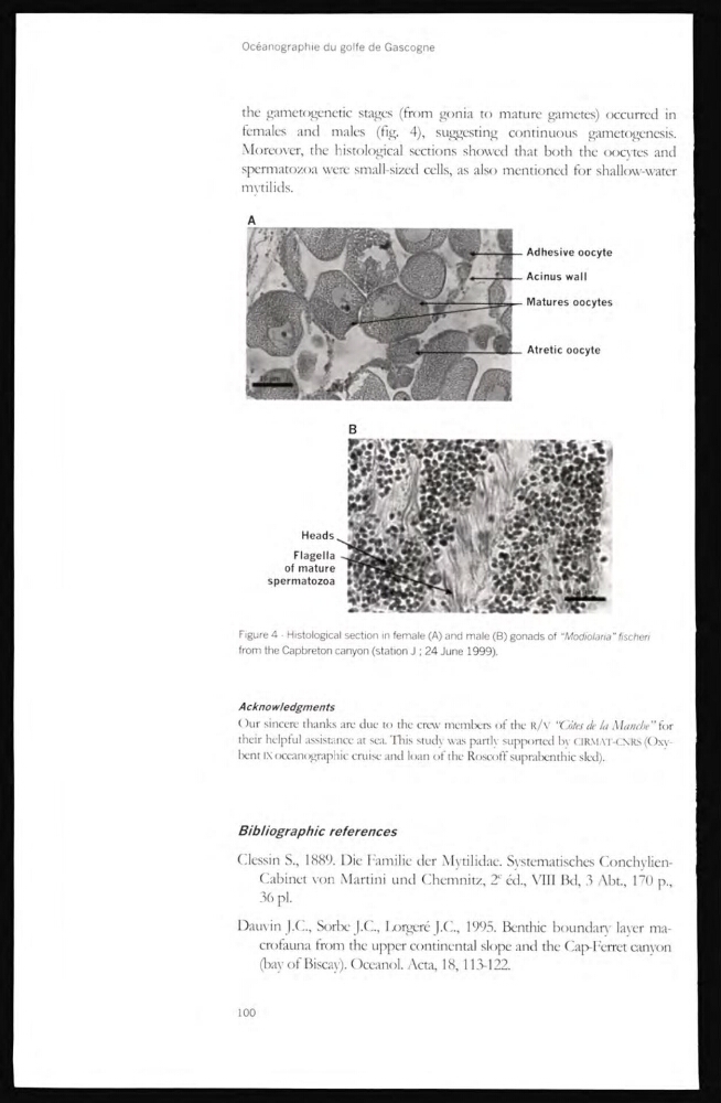
the gametogenetic stages (from gonia to mature gametes) occurred in
females and males (fig. 4), suggesting continuous gametogenesis.
Moreover, the histological sections showed that both the oocytes and
spermatozoa were small-sized cells, as also mentioned for shallow-water
mytilids.
Adhesive oocyte
Acinus wall
Matures oocytes
Atretic oocyte
Heads
Flagella
of mature
spermatozoa
Figure 4 - Histological section in female (A) and male (B) gonads of "Modiolana" fischeri
from the Capbreton canyon (station J ; 24 June 1999).
Acknowledgments
Our sincerc thanks are duc to thc crew mcmbcrs of thc R/V “Côtes de la Manche” for
their helpful assistancc at sca. This study was partly supported bv CIRMAT-CNRS (Oxybcnt IX occanographic cruise and loan of thc Roscoff suprabenthic sled).
Bibliographic references
Clessin S., 1889. Die Familie der Mytilidae. Systemadsches ConchylienCabinet von Martini und Chemnitz, 2
e éd., VIII Bd, 3 Abt., 170 p.,
36 pl.
Damdn J.C., SorbeJ.C., Ixirgeré J.C., 1995. Benthic boundary layer macrofauna from the upper continental slope and the Cap-Ferret canyon
(bay ot Biscay). Oceanol. Acta, 18,113-122.
100
females and males (fig. 4), suggesting continuous gametogenesis.
Moreover, the histological sections showed that both the oocytes and
spermatozoa were small-sized cells, as also mentioned for shallow-water
mytilids.
Adhesive oocyte
Acinus wall
Matures oocytes
Atretic oocyte
Heads
Flagella
of mature
spermatozoa
Figure 4 - Histological section in female (A) and male (B) gonads of "Modiolana" fischeri
from the Capbreton canyon (station J ; 24 June 1999).
Acknowledgments
Our sincerc thanks are duc to thc crew mcmbcrs of thc R/V “Côtes de la Manche” for
their helpful assistancc at sca. This study was partly supported bv CIRMAT-CNRS (Oxybcnt IX occanographic cruise and loan of thc Roscoff suprabenthic sled).
Bibliographic references
Clessin S., 1889. Die Familie der Mytilidae. Systemadsches ConchylienCabinet von Martini und Chemnitz, 2
e éd., VIII Bd, 3 Abt., 170 p.,
36 pl.
Damdn J.C., SorbeJ.C., Ixirgeré J.C., 1995. Benthic boundary layer macrofauna from the upper continental slope and the Cap-Ferret canyon
(bay ot Biscay). Oceanol. Acta, 18,113-122.
100