

2. ELECTROPHYSIOLOGY OF THE RETINA
100- -
g
a,
u
3
-
+
.- -
Q
-
E
50a, m
-
c 0
a
-
m
a
(
L
-
45
o , , , , , , , , , , , , , , , , , ,
1
.
1
'
1
1
4
400
500
600
700
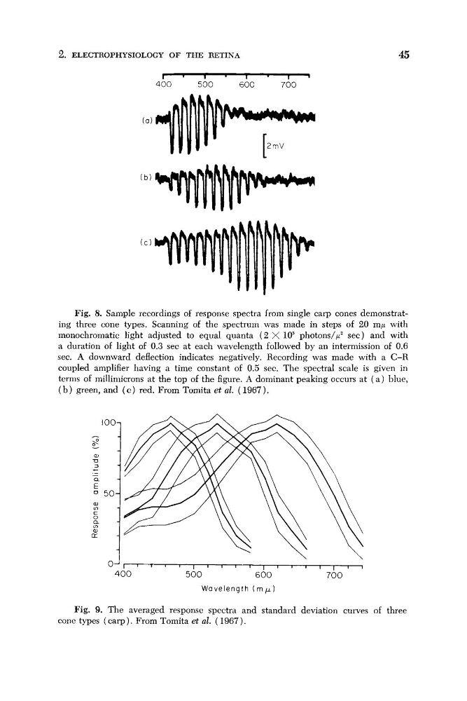
Fig. 8. Sample recordings of response spectra from single carp cones demonstrating three cone types. Scanning of the spectrum was made in steps of 20 mp with
monochromatic light adjusted to equal quanta ( 2 X lo5 photons/$ sec) and with
a duration of light of 0.3 sec at each wavelength followed by an intermission of 0.6
sec. A downward deflection indicates negatively. Recording was made with a C-R
coupled amplifier having a time constant of 0.5 sec. The spectral scale is given in
terms of millimicrons at the top of the figure. A dominant peaking occurs at ( a ) blue,
( b ) green, and ( c ) red. From Tomita et al. ( 1967).
Fig. 9. The averaged response spectra and standard deviation curves of three
cone types (carp). From Tomita et al. (1967).
100- -
g
a,
u
3
-
+
.- -
Q
-
E
50a, m
-
c 0
a
-
m
a
(
L
-
45
o , , , , , , , , , , , , , , , , , ,
1
.
1
'
1
1
4
400
500
600
700
Fig. 8. Sample recordings of response spectra from single carp cones demonstrating three cone types. Scanning of the spectrum was made in steps of 20 mp with
monochromatic light adjusted to equal quanta ( 2 X lo5 photons/$ sec) and with
a duration of light of 0.3 sec at each wavelength followed by an intermission of 0.6
sec. A downward deflection indicates negatively. Recording was made with a C-R
coupled amplifier having a time constant of 0.5 sec. The spectral scale is given in
terms of millimicrons at the top of the figure. A dominant peaking occurs at ( a ) blue,
( b ) green, and ( c ) red. From Tomita et al. ( 1967).
Fig. 9. The averaged response spectra and standard deviation curves of three
cone types (carp). From Tomita et al. (1967).