

1 1. ELECTRORECEPTION
525
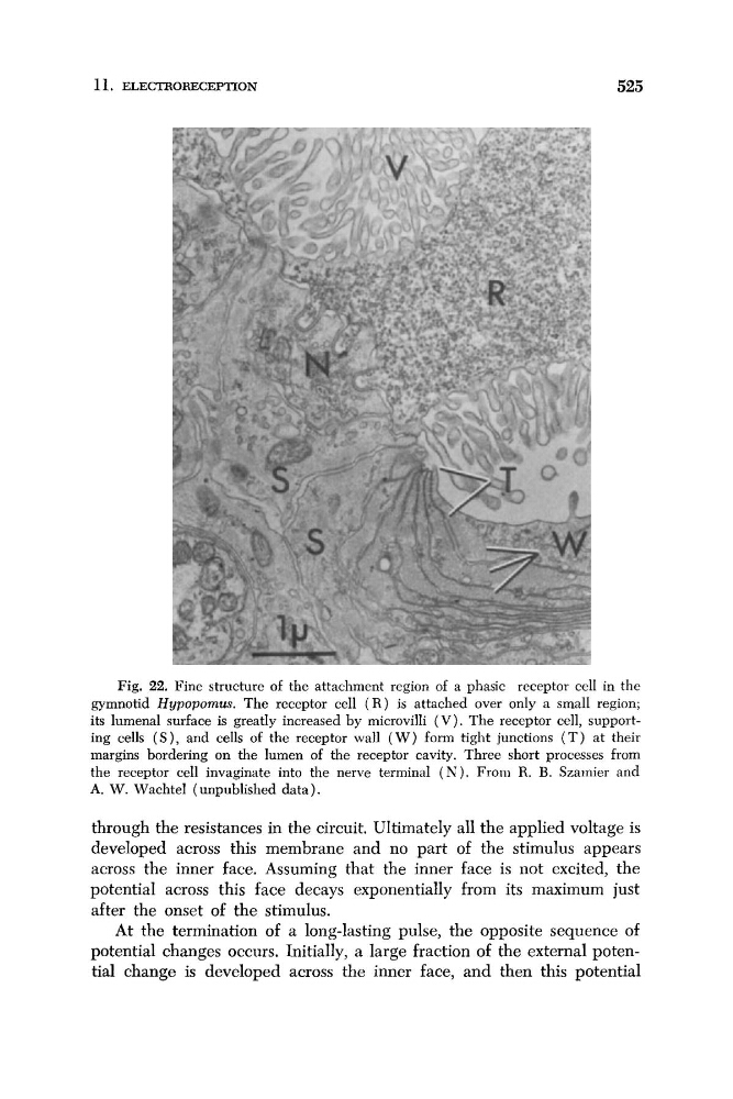
Fig. 22. Fine structure of the attachment region of a phasic receptor cell in the
gymnotid Hypopomus. The receptor cell ( R ) is attached over only a small region;
its lumenal surface is greatly increased by microvilli ( V ) . The receptor cell, supporting cells ( S ) , and cells of the receptor wall ( W ) form tight junctions ( T ) at their
margins bordering on the lumen of the receptor cavity. Three short processes from
the receptor cell invaginate into the nerve terminal ( N ) . From R. B. Szamier and
A. W. Wachtel (unpublished data).
through the resistances in the circuit. Ultimately all the applied voltage is
developed across this membrane and no part of the stimulus appears
across the inner face. Assuming that the inner face is not excited, the
potential across this face decays exponentially from its maximum just
after the onset of the stimulus.
At the termination of a long-lasting pulse, the opposite sequence of
potential changes occurs. Initially, a large fraction of the external potential change is developed across the inner face, and then this potential
525
Fig. 22. Fine structure of the attachment region of a phasic receptor cell in the
gymnotid Hypopomus. The receptor cell ( R ) is attached over only a small region;
its lumenal surface is greatly increased by microvilli ( V ) . The receptor cell, supporting cells ( S ) , and cells of the receptor wall ( W ) form tight junctions ( T ) at their
margins bordering on the lumen of the receptor cavity. Three short processes from
the receptor cell invaginate into the nerve terminal ( N ) . From R. B. Szamier and
A. W. Wachtel (unpublished data).
through the resistances in the circuit. Ultimately all the applied voltage is
developed across this membrane and no part of the stimulus appears
across the inner face. Assuming that the inner face is not excited, the
potential across this face decays exponentially from its maximum just
after the onset of the stimulus.
At the termination of a long-lasting pulse, the opposite sequence of
potential changes occurs. Initially, a large fraction of the external potential change is developed across the inner face, and then this potential