

522
M. V. L. BENNETT
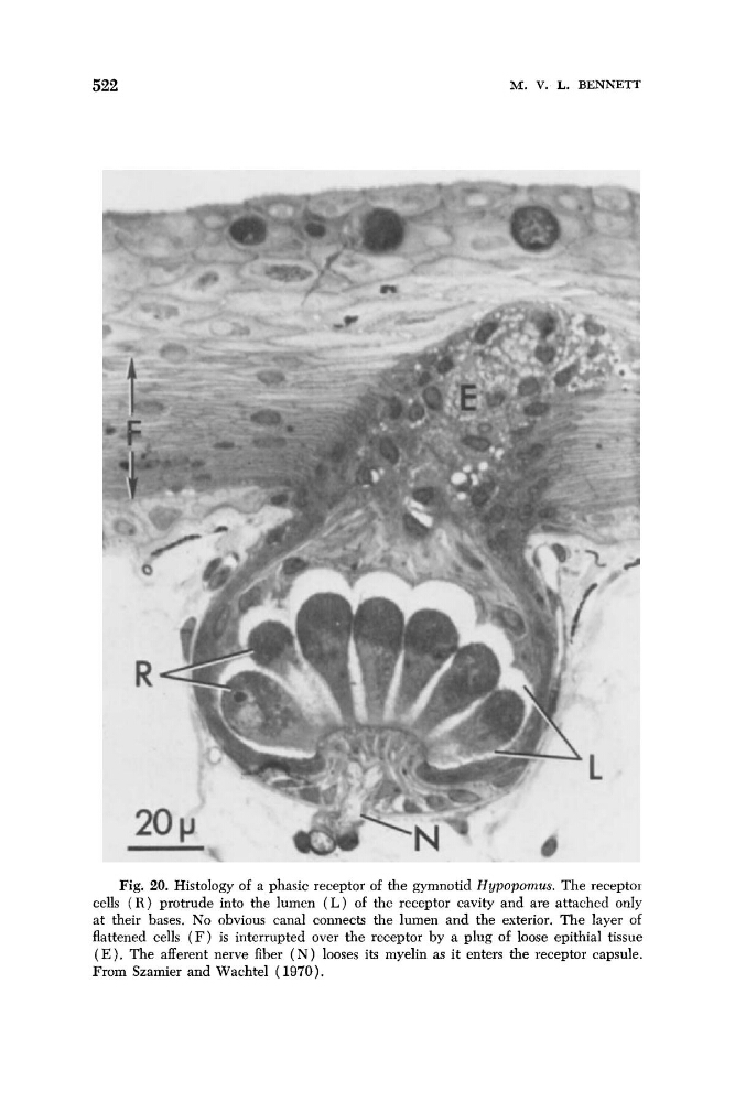
Fig. 20. Histology of a phasic receptor of the gymnotid Hypopornus. The receptoi
cells ( R ) protrude into the lumen ( L ) of the receptor cavity and are attached only
at their bases. No obvious canal connects the lumen and the exterior. The layer of
flattened cells ( F ) is interrupted over the receptor by a plug of loose epithial tissue
( E ) . The afferent nerve fiber ( N ) looses its myelin as it enters the receptor capsule.
From Szamier and Wachtel ( 1970).
M. V. L. BENNETT
Fig. 20. Histology of a phasic receptor of the gymnotid Hypopornus. The receptoi
cells ( R ) protrude into the lumen ( L ) of the receptor cavity and are attached only
at their bases. No obvious canal connects the lumen and the exterior. The layer of
flattened cells ( F ) is interrupted over the receptor by a plug of loose epithial tissue
( E ) . The afferent nerve fiber ( N ) looses its myelin as it enters the receptor capsule.
From Szamier and Wachtel ( 1970).