

1 1. ELECTRORECEPTION
521
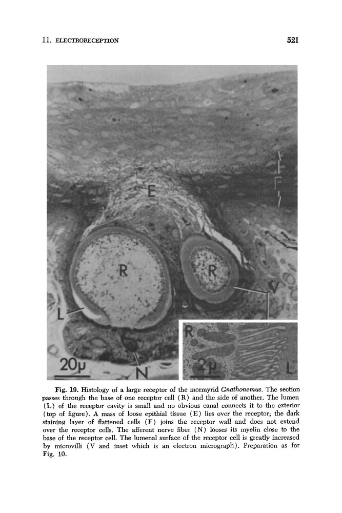
Fig. 19. Histology of a large receptor of the mormyrid Gnathonemus. The section
passes through the base of one receptor cell ( R ) and the side of another. The lumen
( L ) of the receptor cavity is small and no obvious canal connects it to the exterior
(top of figure). A mass of loose epithial tissue ( E ) lies over the receptor; the dark
staining layer of flattened cells ( F ) joins the receptor wall and does not extend
over the receptor cells. The afferent nerve fiber ( N ) looses its myelin close to the
base of the receptor cell. The lumenal surface of the receptor cell is greatly increased
by microvilli ( V and inset which is an electron micrograph). Preparation as for
Fig. 10.
521
Fig. 19. Histology of a large receptor of the mormyrid Gnathonemus. The section
passes through the base of one receptor cell ( R ) and the side of another. The lumen
( L ) of the receptor cavity is small and no obvious canal connects it to the exterior
(top of figure). A mass of loose epithial tissue ( E ) lies over the receptor; the dark
staining layer of flattened cells ( F ) joins the receptor wall and does not extend
over the receptor cells. The afferent nerve fiber ( N ) looses its myelin close to the
base of the receptor cell. The lumenal surface of the receptor cell is greatly increased
by microvilli ( V and inset which is an electron micrograph). Preparation as for
Fig. 10.