

11. ELECTRORECEPTION
505
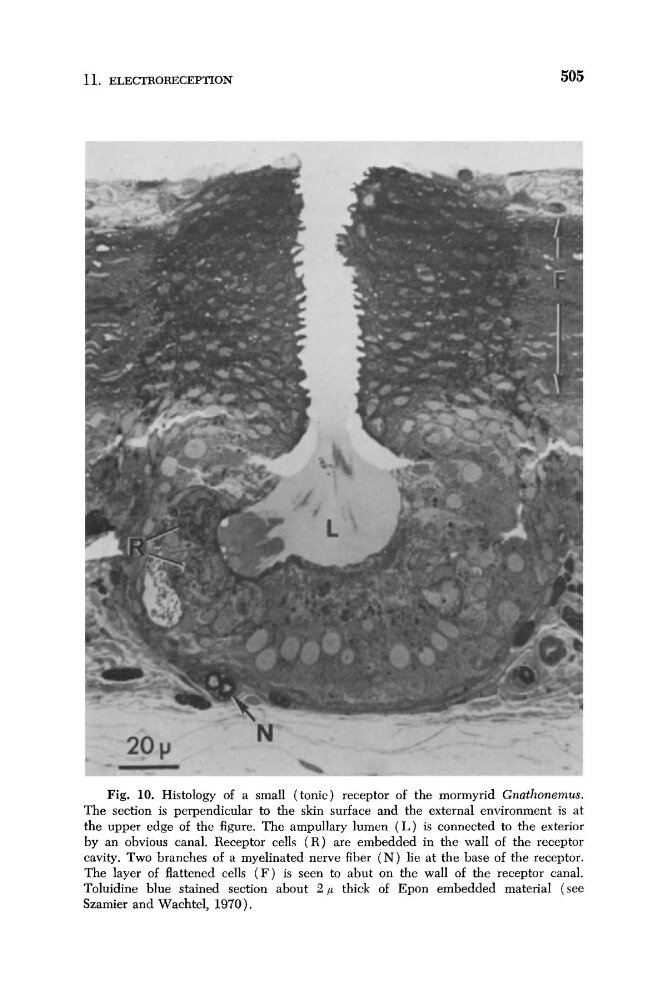
Fig. 10. Histology of a small (tonic) receptor of the mormyrid Gnathonemus.
The section is perpendicular to the skin surface and the external environment is at
the upper edge of the figure. The ampullary lumen ( L ) is connected to the exterior
by an obvious canal. Receptor cells ( R ) are embedded in the wall of the receptor
cavity. Two branches of a myelinated nerve fiber ( N ) lie at the base of the receptor.
The layer of flattened cells ( F ) is seen to abut on the wall of the receptor canal.
Toluidine blue stained section about 2 p thick of Epon embedded material (see
Szamier and Wachtel, 1970).
505
Fig. 10. Histology of a small (tonic) receptor of the mormyrid Gnathonemus.
The section is perpendicular to the skin surface and the external environment is at
the upper edge of the figure. The ampullary lumen ( L ) is connected to the exterior
by an obvious canal. Receptor cells ( R ) are embedded in the wall of the receptor
cavity. Two branches of a myelinated nerve fiber ( N ) lie at the base of the receptor.
The layer of flattened cells ( F ) is seen to abut on the wall of the receptor canal.
Toluidine blue stained section about 2 p thick of Epon embedded material (see
Szamier and Wachtel, 1970).