

11. ELECTRORECEPTION
501
TR H Y O M A N D VII
R M A N D EX1 VENTR VII
R M A N O I N T VII
R M A N O EX1 DORS VII
(C)
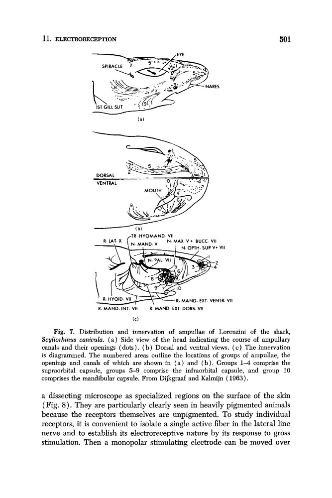
Fig. 7. Distribution and innervation of ampullae of Lorenzini of the shark,
Scyliorhinus cunicula. ( a ) Side view of the head indicating the course of ampullary
canals and their openings (dots). ( b ) Dorsal and ventral views. ( c ) The innervation
is diagrammed. The numbered areas outline the locations of groups of ampullae, the
openings and canals of which are shown in ( a ) and ( b ) . Groups 1 4 comprise the
supraorbital capsule, groups 5-9 comprise the infraorbital capsule, and group 10
comprises the mandibular capsule. From Dijkgraaf and Kalmijn ( 1963).
a dissecting microscope as specialized regions on the surface of the skin
( Fig. 8 ) . They are particularly clearly seen in heavily pigmented animals
because the receptors themselves are unpigmented. To study individual
receptors, it is convenient to isolate a single active fiber in the lateral line
nerve and to establish its electroreceptive nature by its response to gross
stimulation. Then a monopolar stimulating electrode can be moved over
501
TR H Y O M A N D VII
R M A N D EX1 VENTR VII
R M A N O I N T VII
R M A N O EX1 DORS VII
(C)
Fig. 7. Distribution and innervation of ampullae of Lorenzini of the shark,
Scyliorhinus cunicula. ( a ) Side view of the head indicating the course of ampullary
canals and their openings (dots). ( b ) Dorsal and ventral views. ( c ) The innervation
is diagrammed. The numbered areas outline the locations of groups of ampullae, the
openings and canals of which are shown in ( a ) and ( b ) . Groups 1 4 comprise the
supraorbital capsule, groups 5-9 comprise the infraorbital capsule, and group 10
comprises the mandibular capsule. From Dijkgraaf and Kalmijn ( 1963).
a dissecting microscope as specialized regions on the surface of the skin
( Fig. 8 ) . They are particularly clearly seen in heavily pigmented animals
because the receptors themselves are unpigmented. To study individual
receptors, it is convenient to isolate a single active fiber in the lateral line
nerve and to establish its electroreceptive nature by its response to gross
stimulation. Then a monopolar stimulating electrode can be moved over