

498
M. V. L. BENNETT
DORSAL BRANCH OF POSTERIOR
BORDER OF
LINE NERVE
E LECTROREChOR
REGIONS
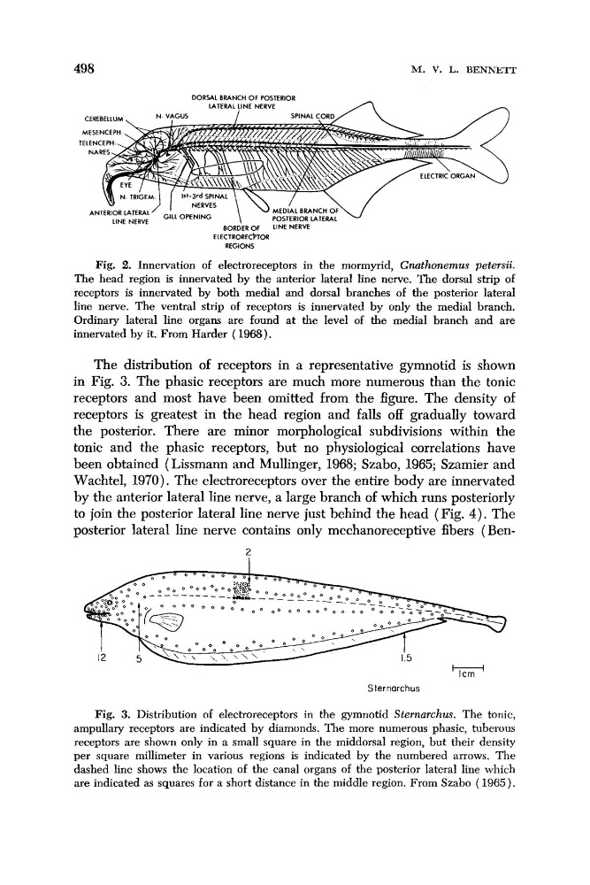
Fig. 2. Innervation of electroreceptors in the mormyrid, Gnathonernus petersii.
The head region is innervated by the anterior lateral line nerve. The dorsal strip of
receptors is innervated by both medial and dorsal branches of the posterior lateral
line nerve. The ventral strip of receptors is innervated by only the medial branch.
Ordinary lateral line organs are found at the level of the medial branch and are
innervated by it. From Harder ( 1968).
The distribution of receptors in a representative gymnotid is shown
in Fig. 3. The phasic receptors are much more numerous than the tonic
receptors and most have been omitted from the figure. The density of
receptors is greatest in the head region and falls off gradually toward
the posterior. There are minor morphological subdivisions within the
tonic and the phasic receptors, but no physiological correlations have
been obtained (Lissmann and Mullinger, 1968; Szabo, 1965; Szamier and
Wachtel, 1970). The electroreceptors over the entire body are innervated
by the anterior lateral line nerve, a large branch of which runs posteriorly
to join the posterior lateral line nerve just behind the head (Fig. 4 ) . The
posterior lateral line nerve contains only mechanoreceptive fibers ( Ben?
S ternarchus
Fig. 3. Distribution of electroreceptors in the gymnotid Sternarchus. The tonic,
ampullary receptors are indicated by diamonds. The more numerous phasic, tuberous
receptors are shown only in a small square in the middorsal region, but their density
per square millimeter in various regions is indicated by the numbered arrows. The
dashed line shows the location of the canal organs of the posterior lateral line which
are indicated as squares for a short distance in the middle region. From Szabo ( 1965).
M. V. L. BENNETT
DORSAL BRANCH OF POSTERIOR
BORDER OF
LINE NERVE
E LECTROREChOR
REGIONS
Fig. 2. Innervation of electroreceptors in the mormyrid, Gnathonernus petersii.
The head region is innervated by the anterior lateral line nerve. The dorsal strip of
receptors is innervated by both medial and dorsal branches of the posterior lateral
line nerve. The ventral strip of receptors is innervated by only the medial branch.
Ordinary lateral line organs are found at the level of the medial branch and are
innervated by it. From Harder ( 1968).
The distribution of receptors in a representative gymnotid is shown
in Fig. 3. The phasic receptors are much more numerous than the tonic
receptors and most have been omitted from the figure. The density of
receptors is greatest in the head region and falls off gradually toward
the posterior. There are minor morphological subdivisions within the
tonic and the phasic receptors, but no physiological correlations have
been obtained (Lissmann and Mullinger, 1968; Szabo, 1965; Szamier and
Wachtel, 1970). The electroreceptors over the entire body are innervated
by the anterior lateral line nerve, a large branch of which runs posteriorly
to join the posterior lateral line nerve just behind the head (Fig. 4 ) . The
posterior lateral line nerve contains only mechanoreceptive fibers ( Ben?
S ternarchus
Fig. 3. Distribution of electroreceptors in the gymnotid Sternarchus. The tonic,
ampullary receptors are indicated by diamonds. The more numerous phasic, tuberous
receptors are shown only in a small square in the middorsal region, but their density
per square millimeter in various regions is indicated by the numbered arrows. The
dashed line shows the location of the canal organs of the posterior lateral line which
are indicated as squares for a short distance in the middle region. From Szabo ( 1965).